The cattle skull is large and massive. It has the shape of a rectangle. The top side of the rectangle has a clear line. It is formed by horny processes and a bone ridge.
The bull's skull is distinguished by a stronger and wider horny ridge. It creates a reliable shield for the brain box. A cow's skull is more elongated. Their front part is elongated. The facial bone of an ox is shorter and stronger. The cranium connects to the cervical spine. The occipital part is massive and strong. It holds the neck muscles. What parts does a cow's skull consist of? What role do they play in the structure of the skeleton?
Cow skull, photo
Fig.1. Cow skull on the left side. Bone boundaries are shown.
1—nasal bone; 2— premaxillary bone; 3— lacrimal bone; 4—upper jaw; 3—zygomatic bone; 6—frontal bone; 7— parietal bone; 8—temporal bone scales; 8′—petrous bone; 9—occipital bone; 10—frontonasal suture; 11— lacrimal suture; 12— lacrimal suture; 13— lacrimal zygomatic suture; 14—incisor-maxillary suture; 15— lacrimal suture; 16— zygomaticomaxillary suture; 17, 18—frontal wedge-shaped suture; 19—sphenoparietal suture; 20—frontozygomatic suture; 21— lower jaw; 22—temporomygomatic suture; 23—frontoparietal suture; 24—temporoparietal suture; 25—occipital-mastoid suture.
Fig.2. Sagittal section of a cow's skull.
1—frontal bone; 2— parietal bone; 3—temporal bone (3a rocky part, 3b squamous part, 3c tympanic part); 4—occipital bone; 5—sphenoid bone; 6—ethmoid bone (6a dorsal concha, 6b perpendicular plate, 6c middle concha, 6d labyrinth of the ethmoid bone); 7—ventral shell; 8—nasal bone; 9— premaxillary bone; 10—upper jaw; 11— opener; 12— palatine bone; 13— pterygoid bone; 14— lower jaw; 15—mandibular foramen; 16—frontoparietal suture; 17—frontal wedge-shaped suture; 18—temporoparietal suture; 19—temporal wedge-shaped suture; 20—occipital-parietal suture; 21— occipital-mastoid suture; 22—occipital-sphenoid fusion; 23— pterygopalatine suture; 24—frontonasal suture; 25—transverse palatal suture; 26—incisor-nasal suture; 27—incisor-maxillary suture.
Fig.3. Cow skull from the dorsal surface.
1—frontal bone; 2— parietal bone; 3—zygomatic bone; 4— lacrimal bone; 5—nasal bone; 6—upper jaw; 7— premaxillary bone; 8—temporal bone; 9—frontoparietal suture; 10—frontal suture; 11— lacrimal suture; 12—frontonasal suture; 13— lacrimal zygomatic suture; 14—maxillozygomatic suture; 15— lacrimal suture; 16— lacrimal suture; 17—nasomaxillary suture; 18—incisor-maxillary suture.
Fig.4. Cow skull from the ventral surface.
1—occipital bone; 2—frontal bone; 2′—horny process; 3—temporal bone scales; 3′—tympanic part of the temporal bone; 4—body of the wedge-shaped host; 4′—temporal wing; 4″—orbital wing; 5— opener; 6— pterygoid bone; 7—zygomatic bone; 8—upper jaw; 8′—its palatine process; 9— palatine bone; 10— lacrimal bone; 11— premaxillary bone; 12—parietal bone; 13— occipital-mastoid suture; 14—occipital-sphenoid fusion; 15—sphenoid-temporal suture; 16—sphenoid-frontal suture; 17—temporomygomatic suture; 18— lacrimal suture; 19— zygomaticomaxillary suture; 20—transverse palatal suture; 21—median palatal suture; 22—occipital-parietal suture; 23—incisor-maxillary suture; 24—palatal incisal suture.
Head
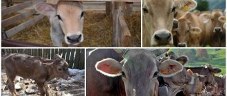
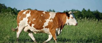
Cows are massive, large domestic animals. And this is easy to determine by looking only at the cow’s head itself. They have a wide, large muzzle with pronounced features characteristic of this species. In particular, a wide flat forehead, a short lower part of the face, well-defined brow ridges, large ears directed to the sides.
A distinctive feature of all cows is their large, expressive eyes. They say that the sky is drowning in these eyes. Another important detail of the head is strong occipital muscles. They help the animal keep its muzzle down for a long time. If I may say so, then most of the time animals keep their heads down because they eat a lot.
Scull
Probably many people have seen the skull of a cow in pictures or in films. These are very strong bones that can withstand a lot of pressure. Unlike deer or elk, neither female nor male cattle ever shed their antlers (if they have them). The bone itself is divided into two halves, which have their own significance for the animal: the brain and the facial.
As its name suggests, the medulla forms a kind of container for the brain. The facial one is responsible for the front part of the cow’s muzzle. This includes the eye sockets, nasal and oral cavities. When a calf is born, both parts of the skull are almost the same size. However, with age, the facial part becomes larger and begins to dominate in size over the brain.
Cow skull: 1-occipital part; 2-facial tubercle; 3-parietal bone; 4-piscus bone; 5-frontal bone; 6-maxillary bone; 7-premaxillary bone; 8-nose bone; 9-lacrimal bone; 10-zygomatic bone; 11-lower jaw; 12-socket; 13-angle of the lower jaw.
In addition to the main two parts, the animal’s head can also be divided into paired and unpaired bones. In total, the cow's skull has 13 paired and 7 unpaired bones. For example, the brain part consists of the occipital, sphenoid and interparietal bones. But from the paired ones we can distinguish the frontal, parietal and temporal.
Eyes
Eyes are not only one of the most striking and beautiful features of a cow’s face, but also a very important analyzer. Like other animals, this part of the head has its own structure, in particular, it has an eyeball, auxiliary and protective organs. The eyeball itself consists of three membranes: reticular, vascular and fibrous. The pupil is located in the iris, which belongs to the choroid.
Udder structure
How many stomachs does a cow have: structural features of the digestive system
The udder is the part of the body for which the animal is so valued, therefore, when considering the anatomy of a cow, this organ should not be missed.
Circulation
Most of the udder is penetrated by a huge number of blood vessels. They saturate the organ with oxygen and take away metabolic products. During milk production, blood circulates from one part of the udder to another at a very high speed.
Lymphatic system
The lymphatic system in this part of the body is well developed. It controls the amount of fluid and removes accumulated toxins and impurities. Each part of the udder has its own lymph node, which swells if the animal has mastitis.
Thanks to the nervous system located in the udder, milk formation and instinctive milk production occur. The nerves send corresponding impulses to the brain, to which the animal reacts reflexively.
Milk follicles
Mammary follicle is part of the mammary gland. Its size and functions depend on the lactation period. It contains special ducts through which milk flows through channels and pipes.
The nipple is a muscular fold that has a sphincter at the end that closes the nipple canal. Thanks to it, milk does not spill out of the udder randomly.
Knowledge about the structure of a cow’s skeleton will allow us to understand the basic needs of a living organism, choose the right nutrition, and improve the living conditions of the animal.
Head structure
Having dealt with the structure of the skull itself, let's move on to considering the structure of the head. It contains the horns, organs of vision, taste and hearing. Let's get acquainted with each one separately.
Many cows have horns. The only exceptions are representatives of cattle, in which this trait was specially removed by breeders. In terms of anatomy, horns are rigid bony formations located on the head of an animal.
The size of the horns directly depends on metabolic processes in the body; their growth is accompanied by the formation of rings. Due to an inadequate diet, young individuals may experience a delay in the growth of horny processes.
Important! Newborns do not have horns. At about one month of age, small tubercles begin to be felt, and in two-month-old calves, small horns can be seen with the naked eye.
Organs of vision
Cows have monocular vision due to their symmetrically located eyes on the sides. The visual organs are large in size, framed by fairly long eyelashes, the purpose of which is not to decorate the eyes, but to provide spatial orientation to the animal. The location of the eyes is round-shaped bony sockets, slightly convex on the outside and flattened on the inside.
The visual organs are connected to the brain, just like in humans. The structure of the eyes contains 3 chambers: external, middle and internal. They are protected from the influence of external factors by eyelashes, eyebrows and lacrimal apparatus.
Skeleton of the neck, torso and tail
How many bones does a cow have in total?
In cattle, the bone is compressed dorsoventrally and greatly expands caudally. The handle is massive, cylindrical, the anterior end is raised dorsally; connects to the body of the bone with a joint. The notch for the first pair of ribs is located at the front end of the handle.
Cervical region
In cattle, the vertebrae are massive and short; the heads and pits are well defined; the spinous processes are developed, have thickened ends, and their length increases in the caudal direction. The costal processes are located ventral to the transverse processes and are deflected forward. The ventral crest is present only on the 3rd - 5th vertebrae. On the 6th vertebra the costal process is wide and long.
Thoracic region
In cattle, the bodies of the thoracic vertebrae, due to their small number (13, rarely 14), are longer than wide. Wide, lamellar, with sharp caudal edges, the spinous processes are strongly inclined caudally. The longest process is on the 2nd vertebra. Diaphragmatic vertebra 13th. Sometimes the ends of the spinous processes are thickened, but flattened from front to back. There are lateral vertebral foramina. The costal facets on the transverse processes are saddle-shaped.
Lumbar
Cattle have 6 lumbar vertebrae; the cranial articular processes are equipped with grooved facets, the caudal ones are cylindrical. The vertebral bodies are long, with ventral ridges, narrowed in the middle. The transverse costal processes are set horizontally or even slightly bent dorsally at the ends; their edges are often jagged and sharp; the length of the processes of the 3rd and 4th vertebrae is greatest. The spinous processes are wide and low. The caudal intervertebral notches are deep.
Cattle have 5 vertebrae. The spinous processes are fused into a ridge with a thickened free edge. The wings of the sacrum are compressed from front to back; ear-shaped articular surfaces are directed caudally; cranial articular processes with grooved facets. The median vascular groove runs on the concave pelvic surface of the sacrum. The ventral sacral foramina are extensive. The fusion of the sacral vertebrae occurs at 3-3.5 years.
Tail section
Cattle have a long tail; there are only 18-20 vertebrae in it, but their bodies are significantly elongated in length. The rudiments of the arches are quite well defined. Of the articular processes, only the cranial ones have been preserved, and the transverse processes have taken the form of wide plates curved ventrally. There are hemal arches attached to the cranial ends of the bodies of the first 3-5 vertebrae, but more often there are only their rudiments in the form of tubercles on the first 9-10 vertebrae.
Components of the skeleton
The general features of the skeleton of animals related to cattle are also inherent in cows. There are differences between the sexes. Males have larger body parts and a more massive skeleton than females. In bulls, the frontal region is significantly prominent.
The skeleton of a cow consists of the bones of the skull, neck, torso, limbs and tail.
The skull is followed by:
- vertebrae of the cervical spine;
- thoracic vertebrae and ribs;
- shoulder blade;
- xiphoid cartilage;
- sternum;
- lumbar vertebrae;
- sacrum and ilium;
- Maklok;
- pubic and ischial bones;
- tail.
The forelimbs have bones:
- radial;
- elbow;
- wrist;
- metacarpus;
- sesamoid and fetlock;
- coronoid and hoof.
On the hind limbs are:
- femur and tibia;
- trochanter;
- knee;
- process of the fibula;
- tarsus, calcaneal tubercle and toe.
Skeletal growth
Mastitis in a cow
Newborn animals of different age maturity differ in the proportions of parts and areas of the body: in calves, compared to cows, the ratio of the length of the legs to the height of the chest is 25% greater, and the ratio of the diameter of the chest to the distance between the breasts is 11% greater. The ratio of the width of the frontal region to the length of the head is 8% greater than in cows.
Age of animals, months.
Live weight, kg Material from the site https://wikiwhat.ru
Absolute skeleton mass, kg
Relative skeletal mass, %
https://zoovet.info/vet-knigi/101-anatomiya-zhivotnykh/domashnie-zhivotnye/8212-sravnitelnaya-anatomiya-skeleta-golovy
https://oferme.ru/zhivotnye/korovy/stroenie-5259/
https://mnogo-krolikov.ru/korovy/opisanie-stroeniya-cherepa-korovy.html
https://wikiwhat.ru/%D0%A1%D0%BA%D0%B5%D0%BB%D0%B5%D1%82_%D0%9A%D0%A0%D0%A1
Head injuries in cows
A cow can get a skull injury when falling, fighting with other animals in the herd, or hitting vehicles. Damage to the head is accompanied by significant blood loss. Superficial injuries require veterinary examination.
In the absence of a fracture or crack, the wound surface is simply treated with hydrogen peroxide, furatsilin or potassium permanganate, followed by lubrication with iodine or brilliant green and sealing. If the wound occupies a large area, sutures are required. In this case, the animal is advised to be kept in a stall with daily treatment of the lesion with an antiseptic to avoid the development of a purulent process.
In cases of severe injury, a probe is used to probe the depth. To stop blood loss in such a situation, a ligature is used. The animal is given an antibiotic: Streptocide or Tricillin. If the cow has lost a lot of blood, a blood transfusion procedure is performed. Treatment is prescribed with drugs with a healing effect: Retinol, Panthenol, Vishnevsky ointment.
Bruises of soft tissues are accompanied by the appearance of hematomas. To treat cracks, alcohol-containing solutions are used. Apply something cold to the problem area and apply a tight bandage. You can make a vodka compress. Cold application continues for 3 days. For the next 3 days, you need to apply something warm to the injured area. Anti-inflammatory ointments are used to treat the bruised area.
With a concussion, open cranial injuries, or concussions, cows are often subject to culling. In this case, only very valuable individuals are treated. Concussion is characterized by the appearance of nervous disorders in animals. In addition, there is an increase in pressure inside the skull with the development of an apathetic state, decreased appetite, and spatial disorientation.
Each section of the skull serves as protection for a specific organ or area in the brain. After an injury, you can predict possible health problems for a cow. A head injury can cause loss of hearing, smell, or vision. To make clear forecasts, you need to know how the skull of cows is structured.
What is the head made of?
Now we’ll find out what parts a cow’s head consists of and what differences they have.
Horns
Many cattle have horns. The only exceptions are specially bred breeds in which breeders have removed this trait. From an anatomical point of view, horns are hard bone formations that are located in the head area.
Their growth is directly related to metabolism and is manifested by the appearance of rings. Inadequate feeding of calves can lead to delayed horn growth.
Did you know? Newborn calves do not have horns, after a month you can feel small tubercles, and at 2 months of age the horns are already clearly visible.
Eyes
Cows have monocular vision due to the symmetrical placement of their eyes on the sides of their heads. The eyes are large and framed by rather long eyelashes, which grow not for beauty, but for orientation in space.
The eyes are located in bony sockets and have the following structure: round in shape, slightly convex on the outside and flattened on the inside.
Cattle eyes have the same connection to the brain as humans. They consist of 3 chambers (outer, middle and inner). Protection from mechanical impact is provided by eyelashes, eyebrows and lacrimal apparatus.
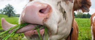
Teeth and tongue
The structure of cow teeth is not the same as that of humans. Incisors and anterior canines are present only on the lower jaw. The upper jaw is covered with durable keratinized epithelium. There are a total of 32 teeth in the mouth.
This jaw structure helps in tearing off strong grass. Retention of food occurs with the help of lips and tongue.
All ruminants swallow food without chewing. After some time, they regurgitate it, grind it with their molars and swallow it again. A newborn calf already has 20 baby teeth. After 1.5 years they are replaced by indigenous ones.
A cow's tongue is a collection of muscle fibers that give it the ability to move.
It performs the following functions:
- food tasting;
- help with swallowing;
- feeling objects;
- body skin care;
- contact with other animals.
Oral cavity
The oral cavity is a complex, well-coordinated organ responsible for the supply of food necessary for the normal functioning of the animal. It is here that food is crushed, moistened with saliva, and then sent down the throat.
Inside, the entire surface of the oral cavity (except for the teeth) is covered with mucous membrane. In some individuals, this shell has pigmentation of varying intensity. The organs of the oral cavity include: cheeks and lips, gums and teeth, hard and soft palate, salivary glands, tongue and tonsils.
The upper lip of the bull is connected to the nose and forms the nasolabial mirror. It can be used to judge the condition of the animal.
The cow's upper jaw remains motionless, while the lower jaw is able to make circular movements when chewing food.
Ears
The bovine hearing system consists of an outer, middle and inner ear. The outer ear detects sounds. It is represented by the auricle with fairly developed muscles, as well as the external auditory canal. The middle ear converts sound vibrations.
This is the eardrum with a chain of auditory bones. The middle ear has a connection to the pharynx. The inner ear is composed of bony and membranous labyrinths.
This is not only the organ of hearing, but also the vestibular apparatus of the animal. So, all the above information allows you to gain complete knowledge about the structural features of the cow’s skull and find out what parts it consists of.
Structure of the cattle skull
The skull of cows consists of two sections: facial and brain. The first section is the location of the visual, olfactory and taste organs, and the second section is the area where the brain is located.
The skull of cows consists of the following bones:
- The back of the head is quite narrow and small. On the outside there are tubercles of muscle tissue formed from small occipital bones. On the sides in the occipital region there are slits, in which there are short jugular processes curved into the middle. In the lower part there are elongated sublingual slits. In the inner part there is an area for a venous vessel. This area connects to the parietal part.
- Top of the head. In young individuals, the parietal region occupies almost the entire cranium. As the animal grows, the crown of the head is replaced by the frontal bone and gradually moves towards the back of the head. The temporal foramina are formed from the parietal bones. The posterior portion of the parietal bone is united with the occipital region, and the parietal plates protect the cerebellum.
- The sphenoid bone, located directly next to the temporal region and orbits. Here are the passages through which the nerve trunks pass, delivering nerves to the organs of vision.
- Frontal bones involved in the formation of the frontal and temporal parts of the skull. The bone tissue of the nose is thick and reaches the eye sockets, although there is a frontal ridge to separate the orbitotemporal region and the nasal bone. At the end of the frontal part there are horny processes.
- Temporal bones connected to the parietal and occipital regions. The temporal bone is the location of the zygomatic ridges, which form tubercles that connect the temples with the cheekbones. In this area there are slits that open to the tympanic cavity.
- Lacrimal bones, connected to the frontal region by zygomatic plates. There is a small gap between the lacrimal bones and the nasal region. In this area of the skull there are pits where the lacrimal sacs are located, from which lacrimal passages in turn emerge leading to the eye sockets.
- The cheekbones are quite massive. These bones participate in the formation of the skeleton of the facial part of the skull.
- Dorsal jaws. The lower jaw row is more massive and wide in comparison with the upper jaw. In the middle there is a jaw tubercle with a toothless edge. The infraorbital fissure is located parallel to the first tooth.
- Incisive bones, formed from plates and externally similar to a roller. The alveoli do not take part in the formation of incisor teeth.
- The palatal bone tissue is highly developed. The hard palate covers a large area of the oral cavity, and the ethmoid bone is involved in the formation of the vertical palate.
- The vomer, presented in the form of a plate, is involved in the connection of the nasal and frontal sinuses, as well as the jaws. This part of the skull is very complex.
- The vertical part, in which 8 alveoli for the incisors are located.
- The hyoid bone, presented in the form of a process, as well as the posterior branches leading to the throat.
In newborn calves, the entire skull is connected by cartilaginous layers, due to which they become mobile, which is important at the birth of a baby. The bones of the skull become hard at approximately 3 years of age, when cartilage tissue is replaced by bone tissue.
Let's get acquainted with some features of the structure of the skull of cows:
- there are horn-like processes on the frontal bones, this area protrudes far towards the back of the head and participates in the formation of the posterior frontal (interhorn) ridge;
- location of the temporal fossae - lateral surfaces of the skull;
- the eye sockets are closed thanks to interconnected processes of the frontal and zygomatic bone tissue;
- the free ends of the nasal bones are divided in two;
- from the slits located above the eye sockets, supraorbital recesses run along the frontal region, directed to the area of the nose and back of the head, and along the frontal region there is a passage to the eye sockets;
- the direction of the long external auditory canals is lateral;
- the incisive bone is poorly developed because it lacks dental sockets;
- the holes are narrow;
- in the naso-caudal direction the cranium is strongly curved: an angle is formed between the base of the cerebral region of the skull and the bony palate;
- in the bone tissue of the mandibular row there are 4 incisive sockets, between them and the sockets where molars grow over time, there are toothless edges;
- the ventral region of the jaws is convex, arched, the processes of the articular tissue are saddle-shaped.