We are accustomed to treating flies as an annoying but harmless nuisance. In fact, even the familiar gray and blue flies are not nearly as harmless as they might seem. Even more dangerous are gadflies, which are also large parasitic flies. All these insects cause diseases collectively called “myiases.” The disease occurs when flies lay larvae in a person and the parasites burrow under the skin. Among this group of insects there are few viviparous insects; the rest lay eggs, and the larvae that emerge from the eggs penetrate the skin. Although there are also “viviparous” flies. Sometimes a person organizes myiasis for himself.
What do a gadfly and its larvae look like?
Gadflies live in almost every corner of the planet; in total there are more than 150 species of insects.
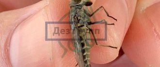
In our country, 60 varieties are registered. Usually, gadflies lay their larvae in the body of animals, less often they get under the skin of a person. Dermatobia Hominis - the “human gadfly” lives in the tropics (Mexico, South America, Argentina). In the temperate climate of Russia, Ukraine and the countries of the former USSR, the insect has not been observed. The adult is a special species of fly measuring up to 20 mm. Dermatobia Hominis looks like a small bumblebee: a shaggy body and a bright orange color. The gadfly has a rather large head with pronounced large eyes, a blue abdomen, and small transparent wings.
Insects living in our latitudes usually have a calmer color: dark brown or coal-black, gray-blue. They prefer livestock as a host, but it happens that when they bite, they also infect humans.
The adult does not feed; the supply of nutrients obtained during the larval development stage is enough for the entire life cycle.
The larva after birth is very small. During the phase, it grows several times, reaching 2 cm. Its body has an oblong teardrop shape. Special hook hairs allow it to attach to the skin of animals or humans.
One adult female can reproduce up to 650 eggs, but only 20% are viable.
A species of dangerous gadfly that lives in southern countries.
Which gadflies lay eggs under human skin?
In Europe, Russia and Asia you can find about 10 species of gadflies (although there are more than 100 of them in the world). All of them do not choose humans as the host for their future offspring. These insects typically lay eggs on cattle or horses. Although cases of infestation of dogs, sheep, cats, rabbits and pigs have also been reported.
But in nature there is a much more dangerous human skin gadfly - Dermatobia hominis . It lives in the regions:
Does this insect bite? No, but its eggs can penetrate humans, where they then turn into parasitic larvae under the influence of heat.
Once in the body, they develop quite quickly. At the same time, their penetration into the body causes inflammation, irritation of the skin, fever, and sometimes vomiting. But the greatest danger occurs when Dermatobia hominis larvae get into the head. Here they can migrate to the eye area. If the larva settles there under the mucous membrane, then there is a high probability of ophthalmomyasis, which in about half of the cases ends in loss of vision.
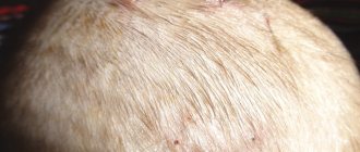
By the way, the larva of a gadfly can live in any part of the human body. Even under the skin on the skull. But most often these parasites choose the limbs, back and armpits.
Types of myiases and flies that cause diseases
Myiasis refers to entomosis, that is, diseases caused by insects. This same group includes, for example, pediculosis. The mechanism for the development of myiasis is simple. Fly larvae penetrate the skin or enter the human body and become parasites, feeding on tissues. Since some larvae can cover a distance of 25–30 cm in a day, the affected area can be very large.
Parasites literally gnaw passages in the skin, muscles, and tissues of internal organs. The damaged areas begin to become inflamed, rot, and the victim experiences severe pain.
Miaz
The most famous flies that lay larvae under the skin of people and animals:
- gadfly (there are more than 150 species);
- fly cabinet;
- blowflies (gray, green, Wohlfart);
- mangrove fly;
- fruit flies.
According to the method of infection, myiases are divided into:
- random;
- obligate;
- optional.
Different types of flies cause different forms of the disease.
Random
This category includes myiases, in which the larvae enter the human body accidentally. Most often this happens while eating. If there are eggs or larvae of flies on the product, the person ingests them with food. In most cases, eggs and maggots dissolve in the caustic gastric juice, but sometimes the larva can enter the intestine, causing intestinal myiasis.
Another example of accidental myiasis is the entry of larvae into the body after a person puts on underwear or uses a towel on which the fly has laid eggs. This can cause genitourinary or ocular myiasis.
Obligate
This type of myiasis is most often found in herbivores (cows, sheep, horses), and is caused by various types of gadflies. Females lay eggs on fur or skin. The hatched larvae penetrate the skin and begin to migrate inside the body, feeding on the flesh of the victim.
Optional
Facultative myiases are caused by flies that lay eggs on carrion. They are unable to pierce the skin, so they lay on open wounds or ulcers on the body. Flies can also lay eggs on mucous membranes. The larva that emerges from the egg begins to eat and burrows deeper into the body. With facultative myiasis, the larvae can even reach the brain.
Myiases in cats and dogs
Myiasis in cats and dogs is not a common occurrence. This is due to the fact that animals carefully lick the affected areas. However, in hard-to-reach places, in weakened animals, this parasitic disease may appear.
The main types of myiases in cats and dogs.
| Types of myiases | Symptoms | Treatment |
| Cutaneous | Anxiety, appearance of wounds. | Remove parasites; treat wounds with antiseptics. |
| Subcutaneous | Loss of appetite, aggressive behavior, festering wounds, itching. In more severe forms - increased body temperature, depression. | Remove parasites; wash wounds with antibiotics; apply Vishnevsky ointment. |
| Intestinal | Depression, vomiting, diarrhea, fly larvae in the stool. | Give a laxative and anthelmintic. |
The larvae are removed from the animal's body under general anesthesia.
Do not ignore the appearance of boils and subcutaneous tubercles. If there is any change in the skin, consult a doctor. The earlier treatment is started, the less harm the parasite will cause to the body.
Larva: what does it look like and reproduce?
The white maggot in a person must go through 3 stages of development. Moreover, at each stage the larva acquires a certain shape.
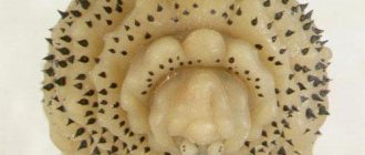
At first these are legless and headless white worms. In one part of the body there is a thickening and three black stripes.
When the larva is in the second stage of development, it increases in size and takes on the shape of a bottle. In the third stage it becomes even larger. Moreover, at each stage, spines and small black dots appear surrounding the thorax of the microorganism.
The larva has 2 posterior spiracles with which it breathes. After being introduced under the human skin, the spiracles remain at the same level with the dermis.
The duration of the initial stage of development is 7 days, after which the larva molts and moves on to the next phase. After 18 days, she sheds again and then the third stage begins.
After a month, the parasite grows into an adult. At the same time, it continues to parasitize the host’s body for about 12 weeks. Next, the gadfly crawls out from under the skin, leaving the person and falling to the ground.
The larva can take liquid food, since its pharynx is adapted to this. It feeds on the fluids and tissues of the host's body, secreting special dermatolytic enzymes that allow it to dissolve solids.
The larva, which has left the human body, pupates in the soil without feeding on anything. After 14-21 days, an adult insect crawls out of it, which after 2-3 minutes becomes ready to fly.
The gadfly has poor eyesight, but very sensitive palps. Thanks to this, females and males quickly find each other and mate.
Features of reproduction
Insects are characterized by sexual dimorphism, that is, it is quite easy to distinguish between a male and a female by appearance. They have different sizes (in many species, females are several times larger than males), differ in color, and the length of their antennae. In some butterfly species, females do not have wings.
Communication between opposite-sex representatives of a species occurs in various ways:
- Using behavioral characteristics.
- Sound and color signals.
- Chemically - by releasing pheromones.
Symptoms of the disease
In the initial stages, especially if the larvae have entered the human skin, there may be no symptoms. Subsequently, as the larvae develop, small pea-sized bumps may form under the skin. Gradually they increase in size and take on the appearance of a local tumor. A telltale sign that you will have to deal with myiasis rather than some other disease is that with myiasis the tumor may move under the skin.
Symptoms may vary depending on the location of the larvae. If you have to deal with cutaneous myiasis, the appearance of a tumor may be accompanied by the formation of small pustules resembling boils.
If the larvae enter the human digestive tract (for example, when they are accidentally consumed with meat products), the course of the disease may be accompanied by symptoms characteristic of ordinary food poisoning. Nausea, vomiting, weakness, lethargy, apathy, sharp pain in the stomach, colitis - these are the most common signs of the disease. This is why it is often confused with a common eating disorder and receives inadequate treatment.
The larvae very quickly infect soft tissues, since they are the basis of the parasites’ diet. The external picture of myiasis differs depending on what type of insect larvae have made their way inside.
https://youtube.com/watch?v=C2OQzgIcq4M
If these are gadfly larvae, most often patients are treated for intestinal diseases. In this case we are talking about a type of myiasis called gastrofilosis. True, this type of disease occurs mainly in artiodactyl animals. The rate of penetration of the larvae into the internal organs is amazing - it can reach up to several tens of centimeters per day. The main goal of the gadfly larvae is to penetrate the stomach and intestines.
If another type of gadfly (the so-called “tropical” gadfly) enters the animal’s body, abscess areas or nodes form in the skin. Their sizes also increase very quickly. Another type of disease carried by tropical flies is cordylobiosis. The clinical picture during the progression of this disease is similar to the previous case. However, in the first case the disease is called “dermatobiasis”, and in the second – “cordylobiosis”.
What harm can the larvae under the skin cause?
The larva of the gadfly contributes to the development of dermatobiasis, which is an obligate myiasis. This disease is characterized by the formation of nodes under the skin, near the worm that has dug into them, which fester and become inflamed.
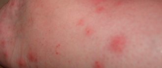
The place where the parasite has entered is similar to a mosquito bite. Over time, this area becomes inflamed, irritated and painful. The diameter of the subcutaneous formation reaches 3 cm; its shape is similar to a boil, from which pus is released.
It is worth noting that the larva can invade any part of the human body - the foot, upper limb and even the head. But most often it lives in the legs, armpits and back.
In some cases, the larva settles under the mucous membrane of the eye, resulting in ophthalmomyasis, which often ends in complete loss of vision. This condition is accompanied by the following symptoms:
- redness;
- lacrimation;
- painful sensations.
Also, the larva of the gadfly can live in the nose. In this case, the patient experiences a headache, discomfort in the nose, his sense of smell worsens and the septum is destroyed. At the same time, the mucous membrane swells, and worms can crawl out of the nose on their own.
In addition, the larvae sometimes parasitize the mucous membranes of the lips, mammary glands and penis. If multiple invasions occur, the formations spread over a large area of skin.
After 12 weeks, the larvae in the human body mature, crawl out of it and pupate.
Dracunculiasis
The disease is caused by subcutaneous parasites - helminths. You can get sick by drinking raw water. It may contain helminth eggs. Once inside, the larvae settle under the skin. It is impossible to notice parasite eggs in water without laboratory tests. And one larva in the human body can lead to death. An adult helminth reaches a size of 100 cm. It can completely occupy the entire volume of the stomach, liver, and block breathing. However, the parasite rarely affects these organs; it prefers the lower limbs.
You can suspect parasites under the skin based on the following symptoms:
- rashes on the skin of the legs;
- bubbles with liquid inside that quickly burst when exposed to water;
- severe itching;
- the presence of compactions in the form of a lump;
- purulent skin lesions.
Treatment involves removing the parasite from the human body. If the larva decides to settle in the internal organs, diagnosis is somewhat difficult. But if you seek help in a timely manner, you can avoid deterioration in your health.
How does a gadfly larva enter the human body?
The gadfly larva can enter the human body in several ways:
- The female lays eggs on the abdomen of blood-sucking insects (mosquitoes, ticks). When a person is bitten by intermediary insects, the eggs land on the person's body. When warmed up, they burst and larvae emerge from them, which get under the skin. The introduction of parasites is practically not noticeable.
- When a human is bitten directly by the female gadfly itself, the larvae enter the wound, after which they fully develop as parasites in the person.
- Hypodermatosis is a disease associated with these parasites. In this case, the larva is acquired tactilely from cattle. It is the countryside and farms in our latitudes that can be considered a potential site of infection. Parasites get under the skin, and they can move along the body, leaving characteristic traces. The larvae usually penetrate the body in areas where the skin is more delicate, for example, on the head, arms and legs, abdomen, neck, and less often they can concentrate on the lips and in the eye.
- Eggs and larvae can also enter internal organs. This occurs when eating animal meat contaminated with gadfly. The gastric parasite is much more dangerous than the subcutaneous larva of the gadfly, since its parasitism can lead to serious disruptions in the functioning of the body.
More complex forms may also occur, when there are several larvae in different areas of the human body.
Is infection possible?
Subcutaneous myiasis occurs due to the laying of eggs of flies or other dipterous insects in wounds or cavities on the human body. In the vast majority of cases, the ears, eyes, long-healing wounds, massive injuries and nasal passages succumb to infection.
In rare cases, subcutaneous myiasis occurs due to the ingestion of fly larvae along with food. This form of the disease is the most difficult to treat.
The following insects can provoke subcutaneous myiasis:
- Sand flea;
- Fly cabinet;
- Wohlfarth fly;
- All types of gadflies.
Please note that the treatment of this pathology must be carried out by a qualified attending physician. This disease is extremely dangerous and can easily lead to death.
Despite the terrible manifestations and serious consequences, treatment of subcutaneous myiasis is carried out with conventional antiparasitic drugs. However, they are taken in larger doses and for much longer. It is extremely difficult to cure subcutaneous myiasis on your own.
In the initial stages, when larvae are visible under the skin, you need to fill them with any oil or Vaseline so that they begin to come out. After this, pick them up with a knife or tweezers.
For subcutaneous myiasis to occur, contact with the pathogen alone is not enough. It is necessary that the insect finds a wound on your body and decides to lay larvae in it. This is quite a rare occurrence for developed countries. Most often, subcutaneous myiasis occurs in residents of hot and humid areas.
For infection, certain conditions must be met:
- Consume water or food contaminated with larvae;
- Ignore personal hygiene rules;
- Swimming in contaminated bodies of water;
- Constant contact with pets;
- Do not treat wounds on the body;
- Touching dead things.
Miaz u
cat
Flies and botflies can lay eggs and larvae on the human body. Larvae can penetrate a person from the ground, mosquitoes, laundry, etc.
Female flies lay eggs in the eyes, ears, nose, wounds of people or inject them subcutaneously; visceral damage is less commonly observed due to accidental ingestion of larvae. For example, members of the genus Gasterophilus, Hypoderma, Dermatobia and Cordylobia affect the skin;
Fannia affects the digestive tract and urinary system; Phonnia and Wohlfahrtia can infect open wounds and ulcers; Oestrus affects the eyes; as well as Cochliomyia penetrate the nasal passages and carry out their invasion.
The possibility of contracting all types of extensive cutaneous myiasis after communicating with an infected person is reduced to zero. This disease occurs due to patient contact with arthropods. It is possible to lay eggs not only in the skin, but also on other individuals (for example, mosquitoes), on things, through the use of which a person can become infected.
Stages of larval development
The larval stage of the gadfly usually lasts 6-10 weeks. After entering the host’s body, the parasite begins to intensively feed on blood, drawing out useful substances. In a few weeks it increases in size tens of times, and the mature larva reaches 2 cm.
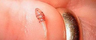
The photo shows a small gadfly larva extracted from a human body.
Having collected the necessary supply of nutrients from the host, the parasite breaks through the skin and crawls out. After this, a new stage of development of the gadfly begins - the pupa. In this phase, the insect arrives for 2-4 weeks, after which it turns into an adult, the life cycle of which is 20 days, the main task of the fly is reproduction.
Diseases caused by protozoa
Leishmaniasis
Leishmaniasis is caused by protozoan single-celled pathogens transmitted by mosquitoes. A person infected with leishmaniasis becomes a reservoir for further spread of the infection. After a mosquito bite, which is the main host organism for leishmania, a person develops cutaneous or visceral leishmaniasis. Cutaneous leishmaniasis appears as deep ulcers or pustules, as well as extensive skin lesions. The mucocutaneous form of the disease leads to significant deformations of the appearance, especially of the face. Swelling of the airways due to leishmaniasis can be fatal.
Leishmaniasis occurs in 90 countries and is a very common disease in Syria, Iran, Afghanistan, Saudi Arabia, Brazil, and Peru.
Main routes of penetration
The female hunts for blood-sucking insects - ticks and mosquitoes. The gadfly lays eggs on their abdomens.
When bloodsuckers land on human skin, the larvae feel warmth. Against this background, they begin to actively hatch from eggs. Then they move on to more suitable “territory”. The implementation takes place within a few seconds.
Note! The invasion is not accompanied by pain or even minor pain syndrome.
Usually a person does not even notice and does not know that a “stranger” is settling in him. The first symptoms appear when the creatures begin to actively develop, parasitizing in an environment favorable to them.
The gadfly prefers to implant its offspring where the skin is thinnest - on the face. Sometimes insect eggs penetrate internal organs. This happens when a person eats meat from infected animals.
The intestinal parasite is much more dangerous than all the others. Against the background of its parasitism, serious gastrointestinal pathologies develop.
Schistosomiasis
The disease is caused by a certain type of helminth that lives in water bodies. You can get sick while swimming in rivers in Africa and Asia. In addition, helminth eggs may be present in drinking water. Subcutaneous parasites initially settle under the layer of the epidermis. An allergic reaction appears on the skin in the form of hives and severe itching. The larvae then penetrate the internal organs. They affect the liver, kidneys, and genitourinary organs. At night, the patient experiences attacks of fever and increased sweating. When diagnosing the disease, specialists pay attention to an enlarged liver and deformed kidneys. Treatment is carried out with antimony drugs. To avoid getting sick with schistosomiasis, it is recommended to drink only boiled water and not swim in local rivers outside the resort area.
What are the signs of infection?
At the initial stage, there are no specific symptoms indicating that a terrible worm has settled in the human body. A slight swelling appears at the site of “foreign” penetration. The shade of the formed tubercle is pinkish. A gadfly bite can easily be confused with a mosquito bite or any other fly.
Then the following signs appear:
- development of the inflammatory process,
- the appearance of painful sensations. They progress and can be quite painful,
- the appearance of reddish or bluish swelling.
The next stage is the formation of an abscess. It can be confused with a large boil. After some time, the abscess opens. Then purulent contents flow out of the wound. There can be a lot of it - it all depends on the size of the abscess.
The patient's health deteriorates significantly. The person feels sick, vomits, and becomes very dizzy. In the most severe cases, the patient loses consciousness.
An acute feverish state is combined with noticeable muscle pain. At the site of the inflammatory focus, under the skin, the movement of something living is felt.
When the pest gets into the nose, the person complains of severe headaches. The sense of smell becomes much worse. The patient complains of discomfort and pain in the nose. The mucous membranes swell, the larvae escape through the nasal passages.
If the gadfly has penetrated the organs of vision, then as they advance, irritation of the mucous membrane occurs. There is an increase in eye pressure. Some people experience involuntary bleeding. Sometimes bloody impurities appear in tears. A person complains that his eyes hurt a lot. At first, the painful syndrome in the eyeballs is not very strong, and many people confuse it with the discomfort that occurs when intraocular pressure increases.
Establishing a diagnosis
If a person suspects he is infected, he should seek help from a good parasitologist or infectious disease specialist.
The doctor sends the patient for a blood test, which determines the amount of antibodies. The specialist undertakes to find out from his client whether he has been to African countries or other places where the terrible gadfly lives.
A visual inspection is required. If infection occurs, an abscess with a fairly large hole forms on the cover.
Inspection is carried out using a special magnifying glass.
Surgery
Surgery must be performed under as sterile conditions as possible. The affected area is carefully treated with an antiseptic drug. It is allowed to use potassium permanganate, or iodine.
In order to block air access to the parasite, a small amount of sterile oil is dripped onto the skin. When the parasite feels threatened, it will begin to independently escape from the body of its host. The doctor can “help” him, “armed” with a special clamp or tweezers.
Then the specialist carefully treats the wound and makes a sterile dressing. In order to relieve inflammation, the patient is prescribed a course of antibiotics.
You should not try to open the abscess and pull out the larva yourself - this can lead to catastrophic consequences. At best, the inflammation recurs; at worst, the patient will develop blood poisoning.
When the wound heals, the use of special oils, creams and ointments is allowed.
What harm is done to the body
The degree of adverse effects depends on where exactly the insects that have entered the body are located. There is a violation of the general state of health. Dysfunction of all vital organs occurs. Against the background of poisoning of the body with waste products of the larvae, the development of powerful intoxication is observed.
The greatest danger is posed by larvae that are localized in:
- intestines,
- ENT organs,
- eyes,
- stomach.
Important! If the larva is not detected in time, it can easily penetrate the vitreous body.
It’s worse if the pest gets into the anterior chamber of the eyeball. As a result, the person partially loses his vision. In the worst cases, complete blindness develops.
It is possible to defeat parasites!
— Reliable and safe removal of parasites in 21 days!
- The composition includes only natural ingredients;
- Does not cause side effects;
- Absolutely safe;
- Protects the liver, heart, lungs, stomach, skin from parasites;
- Removes waste products of parasites from the body.
- Effectively destroys most types of helminths in 21 days.
There is now a preferential program for free packaging. Read .
Hello, readers of the site about parasites Noparasites.ru. My name is Alexander Lignum. I am the author of this site. I am 23 years old, I am a 5th year student at the Kemerovo State Medical Institute. Specialization "Parasitologist". More about the author>>
Search for cures for parasites
Other tests
Classification of disease types and signs of invasion
Depending on which tissues or organs are affected, myiases are divided into:
- subcutaneous;
- intestinal;
- genitourinary;
- ophthalmic;
- nasal;
- oral;
- ear
Each form has its own symptoms. What is common is a deterioration in well-being. A person experiences itching and pain at the site of penetration of the larva. Weakness, nausea, dizziness, and headache appear. Possible increase in temperature and fever.
Subcutaneous
The fly does not lay larvae under the skin, since the ovipositor is not capable of damaging the skin. The laying is done either on the surface of the skin or in existing damage. The hatched larvae independently penetrate the epidermal layer. This is how subcutaneous myiasis develops. The main symptoms are red-purple blisters on the skin (sometimes with a hole in the center). When the larvae move, the victim experiences pain and itching. Parasites can be located shallowly, in the upper layer of the skin, or penetrate deeper into the connective tissues and further into the muscles and tendons.
Intestinal or cavitary
Intestinal myiasis can be caused by almost any type of insect, even ordinary house flies. The larvae enter the human body through the gastrointestinal tract along with food. Most often, the larva dies after a few days and is excreted in feces or vomit. But sometimes (most often with low acidity) the larvae begin to develop in the intestine, penetrating its mucous membrane.
Symptoms of intestinal myiasis:
- pain in the abdomen and anus;
- nausea, vomiting;
- diarrhea;
- weight loss.
In the advanced stage, internal bleeding appears.
Urogenital
This type of myiasis appears due to the fact that fly larvae penetrate the urethra. Most often this happens after a person puts on underwear, on the surface of which the female has laid eggs. After the parasites penetrate the human body, their vital activity becomes the cause of inflammatory processes in the genitals or bladder. A common symptom is difficulty urinating, pain in the urinary canal.
Ophthalmic
Ophthalmomyasis is one of the most severe types of disease. It occurs after fly eggs or larvae enter a person's eye. Parasites penetrate into the thickness of the conjunctiva, into the mucous membrane, or even into the eyeball. This leads to pain, decreased visual acuity, and in advanced cases can cause complete blindness.
Nasal or nasal
This form of myiasis is quite rare in humans. Most often, wild animals become infected with it. In most cases, the cause of the disease is nasopharyngeal gadfly. These viviparous insects seem to inject larvae into the nostrils or eyes of the victim. The larvae invade the nasal mucosa, and then can move further and even reach the brain. Symptoms of infection:
- pain in the nose;
- increased secretion of mucus;
- frequent sneezing;
- purulent rhinitis.
As the parasites move inside the body, the location of the pain changes.
Oral
Cases where a fly has laid eggs or larvae in a person’s mouth are extremely rare. This is primarily due to the fact that foreign organisms in the oral cavity are very easy to detect. But if infection does occur, inflammation, ulcers, and fistulas appear in the mouth.
Ear shape
Otomiasis develops when a fly lays eggs inside the ear or on the surface of the auricle. You can also become infected if you sleep on linens that have eggs or larvae on them. The larvae hatching from laid eggs penetrate deep into the auricle, causing pain and hearing impairment. In severe cases, the parasites penetrate the brain.
Intestinal myiasis
In addition to these species, intestinal myiasis can be caused by almost any fly. But this type of disease takes its most serious forms when it is infected by fruit flies and cheese flies.
Symptoms:
- irritation and inflammation of the intestinal mucosa;
- abdominal pain;
- diarrhea with larvae;
- exhaustion;
- sharp pain in the anus;
- vomiting with maggots.
On a note!
Adult maggots reach a length of up to 1.5 cm. Treatment is prescribed after diagnosis and is carried out using anthelmintic drugs against nematodes.
How does a larva behave when it enters a human body?
The parasite stays in the host's body for 5 to 10 weeks. During this time, it goes through several stages of development. After 30-40 days, the gadfly larva becomes an adult. But it does not leave the owner's body. She needs to get enough and become even bigger.
In this case, the larva is positioned so as to receive oxygen. It breathes through 2 posterior spiracles, which, after being introduced under the human skin, remain at the same level with the epidermis. Is the place where the parasite has entered noticeable? Yes, at first it looks like a mosquito bite, but after a couple of days swelling and redness appear. A bruise may also form.
While under the skin, the larva intensively feeds on blood and absorbs other useful substances. During the time spent in someone else's body, it increases several times. The parasite has a very good appetite. After all, he must stock up on food for the rest of his life. The fact is that after turning into an imago, the insect no longer requires any food.
Growing in maggots
A good alternative to rotten beef, fish in paper, eggs and bottles is maggot. This is the name for a design for raising larvae, which allows almost no interaction with them and reduces the unpleasant smell of rotting meat to a minimum. It consists of three containers placed on top of each other. The top and bottom have holes in the lid, and the top and middle have holes in the bottom. How it works?
Place a spoiled piece of beef or fish in the first container and place the maggot in a dark place where flies can easily penetrate. The second and third containers are filled with sawdust: they will absorb the smell of rot. After 2-3 days, the flies will leave eggs there, the container can be closed and left in the same place. The hatched larvae will fall through the holes into the second compartment, where the sawdust is. Here they will clear and fall into the third compartment. From there they are taken and fed for fishing. This will take up to 10 days, and contact with meat and maggots will be minimal.
Diagnosis and treatment
Moreover, the disease caused by the larvae of the human gadfly has a name - dermatobiasis. It is characterized by the formation of painful nodes under the skin around the parasite. Such bumps can become inflamed. Often pus comes out of them.
At the same time, the human body reacts sharply to the “stranger,” which leads to the appearance of various allergic reactions. The larva itself also secretes the most harmful substance, hypodermotoxin. As a result, symptoms such as:
- fever;
- vomiting (or attacks of nausea);
- weakness (usually with dizziness);
- muscle pain;
- skin redness;
- itching;
- swelling;
- tearing (if the larvae get into the eyes or mucous membranes).
Of course, if parasites are detected in a timely manner and removed, no life-threatening complications arise. All the unpleasant reactions described above are quickly eliminated by promptly removing the parasite, taking medications correctly and using special ointments.
The main thing is to timely diagnose the disease caused by the vital activity of the larvae. To do this, a blood test is performed to determine the amount of antibodies. If they do not correspond to the norm, and the patient himself has been in places where gadflies are active, then, most likely, an infection has actually occurred.
Sometimes a simple visual examination is sufficient, during which purulent abscesses with a hole are identified on the skin. The doctor can only examine them to find parasites.
Filariasis
When going to warm countries on vacation, it is advisable to study not only local attractions and paradises, but also the diseases that may lie in wait there. Local insect bites cause skin damage, discomfort, and unbearable itching. But this is far from the most
Filariasis
scary. The bite of one insect can infect another. This is what happens with filariasis. The danger of the disease lies in the long incubation period. The parasite develops under the skin, lays eggs, and affects internal organs for 7 years. A person does not even know about the existence of parasites under the skin. Parasites are common in Africa, South America, and Asia.
Initially, skin lesions appear in the form of urticaria. Feels itchy. As they reproduce
parasite, the amount of waste products increases. Toxins cause malaise, weakness, and fever. If the parasite reaches the eye, vision decreases. After some time, papules, eczema, ulcers, and ulcers appear on the skin. Larvae in the form of worms move under the skin. The patient feels it and in some cases can be seen with the naked eye. A person can only get sick after being bitten by an insect that carries eggs. As a rule, it is a blood-sucking parasite. The disease is not transmitted from a sick person to a healthy person. The disease is diagnosed at a late stage, when the larvae are already formed. They are viewed with special medical devices. Until then, doctors will not be able to make a correct diagnosis. A person will feel relief after removing the parasites from the body and undergoing appropriate treatment.
Consequences
Infection with the subcutaneous botfly leads to the following consequences:
- Cows have a decrease in milk yield by approximately 7%.
- Young animals have growth retardation.
- For the leather industry - the skin of animals that have suffered from hypodermatosis has holes, which spoils the raw hides.
- For the meat industry, the capsules in which the larvae developed require removal, due to which a fairly large amount of meat is lost; sometimes, with severe contamination, about 10% of the raw materials have to be cut out.
If signs of infection are detected, animals are slaughtered exclusively in sanitary slaughterhouses.
Demodicosis
The disease is caused by subcutaneous parasites – demodex mites. Localized on the face, settle in the sebaceous glands. Externally, the manifestations of the disease are characterized by the presence of acne. Cosmetics and medications for cleansing the skin do not give the desired result. The entire surface of the face is covered with pits, tubercles, ulcers, and inflammation. It has a very unattractive appearance. You can get sick from a sick person or through his things. Low immunity of the body and unstable hormonal levels contribute to infection. Parasites can live under the skin for a lifetime without proper treatment. A dermatologist is engaged in therapy. Treatment for demodicosis will take a long time. Sometimes this takes about a year.
Prevention
In order to prevent the spread of the bovine botfly, animals must be periodically examined for the presence of fistulas.
- During the period from March to May, it is advisable to carefully palpate the backs and lower backs of cows and horses - this technique will allow you to detect subcutaneous nodules in time.
Important! If nodules are detected, you should immediately contact your veterinarian!
- For preventive purposes, at the end of summer or early autumn, cows and horses are treated with special preparations, the action of which is aimed at destroying the larvae that are in the first stage of development. Moreover, absolutely all livestock are subject to processing, including animals that are the property of individual owners.
- In order to prevent the botfly larvae from penetrating the skin after they emerge from the eggs, during the grazing period it is recommended to graze animals before 10.00 and after 18.00. During the daytime, it is advisable to keep cattle under sheds or indoors.
Preventive measures
In order to avoid the manifestation of myiasis, you should follow some rules:
- Use various insect repellents;
- Carry out strict disinfection of various wounds on animals;
- In order to prevent deep damage, it is necessary to promptly treat any damage to the skin;
- An important role is played by the destruction of pests and their breeding sites;
- Comply with food storage standards;
- It is strictly forbidden to eat foods that have expired;
- Eat only washed vegetables and fruits;
- Drink exclusively purified water;
- Observe personal hygiene standards (washing hands, regularly changing linen, etc.);
- Eliminate bad habits in children from an early age.
There are general recommendations during the treatment period. You should use decoctions of black currant leaves, drink plenty of fennel tea, and decoctions of the medicinal plant celandine. It is recommended to add garlic, cumin, onion and cinnamon as spices to various dishes.
For the skin form of the disease, you should use special ointments made from birch tar or sulfur. For the intestinal form of the disease, it is possible to use enemas with bitter medicinal herbs.
Are gadfly larvae dangerous for pets?
We have found out that humans and cattle can suffer from these parasites. What about our pets? Especially dogs and cats that periodically walk outside. There is a certain risk.
At the same time, infection of dogs and cats occurs not only through contact with intermediary insects. These animals like to lie on the ground or in the sand, where gadflies also often lay their eggs. It turns out that a dog or cat, having run around, lies down to rest, which makes it possible for the larvae to get onto the pet’s body. Also, the most likely places of infection are areas with high vegetation and areas with a fairly large population of rodents.
It is not difficult to notice symptoms of infestation with gadfly larvae in dogs or cats. As a rule, the animal begins to behave passively, coughing, and breathing heavily. He often experiences loss of coordination, fever, and even paralysis of the limbs. If at the same time there are tubercles and bumps on the pet’s skin with an obvious opening for the vital activity of the parasite, then, most likely, this is indeed an infestation by botfly larvae.
It is necessary to promptly seek help from a veterinarian. With a greater degree of probability, “aibolit” will prescribe antiparasitic medications that can neutralize insects and alleviate symptoms. If possible, he will also perform surgical removal of parasites from your pet’s body.
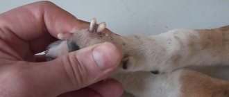
For reference! There is a known case where more than a hundred gadfly larvae were found under the skin of a dog. At the same time, the parasites managed to spread throughout the body. They were on the paws, back, stomach, lips and ears. Only timely medical assistance saved the four-legged animal’s life.
Scabies and scabies mites
Any person, young or old, can get scabies. But the disease is very common among homeless people, disadvantaged members of society. The disease is caused by the scabies mite. A small insect about 1 mm in size. Under the skin, parasites build tunnels, feed, and lay eggs. They crawl to the surface of the skin to reproduce at night. Ticks are highly fertile. Larvae lead the same lifestyle as adults. The incubation period of the disease is about 2 weeks. During this time, the female tick has time to lay eggs, and a new generation is born. Initially, the scabies mite affects the hands - the space between the fingers. Blisters filled with liquid appear, which, after opening, become crusty. The unbearable itching gives no rest. Intensifies in the evening and continues until the morning. It is at this time of day that the scabies mite leads an active lifestyle. In just 1 month of illness, subcutaneous parasites spread throughout the body. The stomach, arms, legs, buttocks, chest, and genitals begin to itch. And in children, skin lesions are visible even on the face.
Currently, scabies can be treated quite quickly and effectively. A special ointment, for example, benzyl benzoate emulsion, is used to treat the entire skin. Clothes and bedding are washed in hot water, then ironed with steam. After 2 days, take a shower and repeat the procedure again. As a result, the scabies mite dies within a week. The disease is cured faster if treatment begins in a timely manner. If left untreated, the scabies mite causes severe skin damage, inflammation, and infection.
Larval therapy - what is it?
Research conducted by scientists has proven that the use of maggots of a certain type, worms in cleansing wound surfaces is a progressive treatment method.
The time spent on cleaning is up to 5-6 days, but the use of traditional methods allows you to achieve a similar result only on the 90th day.
Doctors recommend the widespread use of the technique, for example, in the treatment of methicillin-resistant staphylococcus.
Treatment with maggots is a fairly forgotten old remedy. It was used both in ancient times and during the Second World War, but the advent of antibiotics led to a decrease in the popularity of therapy. Today, the technique is in demand among adherents of alternative therapy in our country, as well as as an adjuvant in European clinics.
The essence of the technique is that maggots eat only dead tissue and do not touch healthy areas. This is what attracted the attention of specialists.
The opinion of professionals is clear: the use of sterile larvae and maggots of flies can significantly speed up the cleaning process, and therefore the healing of wounds.
In addition, there are no problems with drug compatibility, and the risk of wound infection by antibiotic-resistant bacteria is reduced.
What larvae are treated?
Larvae of common flies are used to sterilize and eliminate tissue necrosis. These unattractive worms can work wonders. Maggot larvae, applied to the wound, eat away and eat away the necrosis with such skill that is inaccessible to some surgeons.
However, despite all the positive aspects, not every patient will be able to undergo the course of therapy. Allowing there to be worms in the wound means having fortitude and not suffering from disgust.
The method is simple: sterile maggots are applied to a wound with necrosis and pus. The top crust must then harden so that the worms can eat away the dead flesh.
After all, the wound is opened, the larvae are removed and you can wait for the healing of the flesh.
The principle of action of maggots on the patient’s body
Scientists have experimentally found that the ability of fly larvae to heal wounds is caused by suppression of the body's immune response. A substance secreted by fly larvae when combined with blood serum causes a decrease in protein levels. In some cases, the concentration is reduced by almost 99%.
A detailed study of blood samples showed: mucus components break down complement C3, C4, which leads to suppression of the immune response and faster healing of wounds.
Moreover, the healing process is not burdened by purulent inflammation of healthy tissue, swelling, swelling and focal redness are completely absent - characteristic signs of the onset of an infectious process in healthy tissue.
Fact! The mucous substance does not lose its beneficial qualities even when boiled or kept for a month - this property ensures the effectiveness of larval therapy even in the case of particularly advanced gangrenous, purulent processes.
Today, doctors successfully use the antibiotic seraticin, isolated from the mucus of maggots, and used to treat trophic ulcers and bedsores.
Treatment technique
To get the right maggots, the flies are kept in a sterile, enclosed area where they can lay their larvae. The worms are then placed in bags and only then can the “live medicine” be used. The effects of worms on the human body are as follows:
- sterilization of focal wounds;
- stimulation of healing;
- cleansing by eating necrotic areas;
- the secreted substance allantoin stimulates the healing process.
Fact! Allantoin, secreted from the urea of the larvae, is also found in cow urine. That is why in villages they still wash wounds with evaporated cattle urine.
It should be noted that seraticin, an antibiotic contained in the mucus of worms, resists 12 strains of methicillin-resistant Staphylococcus aureus, destroys E. coli and bacteria that cause pseudomembrane colitis.
Larval therapy includes several mandatory stages:
- breeding larvae of a certain type of fly (green fly, blowfly);
- obtaining eggs with their subsequent washing and sterilization;
- hatching larvae;
- placing worms in the wound;
- opening the wound and removing maggots.
Before placing worms into a wound, they are forced to starve, so placing larvae in a wound for more than a day is not recommended. However, sometimes the time of therapeutic effect is calculated individually.
It all depends on the severity, size of the affected area, type of wound and the presence of purulent inflammation. For example, if the lesion is chronic, the bed is covered with disinfected larvae for 4 days.
The procedure can be repeated several times, achieving complete cleaning of the wound bed and a speedy recovery of the patient.
Interesting! Sometimes silky lucilia larvae are used. These worms secrete an enzyme that dissolves dead tissue and then eat the resulting substance. After 2-4 days, individuals grow to a size of 12 mm and stop cleaning the wound. If necessary, they are replaced with a new portion of maggots and therapy is continued.
Unfortunately, larval therapy in its original form causes more rejection than acceptance in the average patient. Not every person can allow worms to appear in a wound, and doctors prefer more conservative methods of healing.
But if the doctor suggests trying this option, you should not refuse - in just 1-2 days the bed of the most advanced wound will be cleared, and the healing process will go much faster.
In this case, you will not have to administer loading doses of antibiotics and other anti-inflammatory medications.
Treatment of myiasis with traditional methods
Folk remedies for the treatment of myiasis are also no less effective. Methods that have been proven over the years are still used today. They can be independent, but to prevent deterioration of the condition and better treatment, traditional medicine must be used in conjunction with medications and under the supervision of a doctor.
Birch tar and sulfur are often used. For 4 tablespoons take 6 grams. sulfur and 3 tablespoons of Vaseline. This remedy is used for skin diseases.
For intestinal pathology, you need to drink tea from black currant and fennel leaves. Onions and garlic are also known for their properties. Cumin and cinnamon are good in the fight. Use celandine tinctures. To use it, take a spoonful of herbs with roots and pour one glass of boiled water. They drink it before breakfast and lunch.
Worms and parasites in a human wound: what are they and how dangerous?
This spectacle is not for the faint of heart. Sometimes it occurs naturally. And sometimes it is created artificially, called larval therapy. What are the dangers? How effective is this treatment method?
Various sources report that a person developed worms in a long-healing wound. Often these are maggots. It’s not pleasant to read about this, but to see how parasites swarm in the human body is something!
Even more surprising is that parasites can serve as “orderlies” and “healers.”
A human wound with worms and parasites - what is it?
If you do not properly treat the rotting lesion, do not change the dressing material in a timely manner, and keep the patient in unfavorable conditions, then maggots appear in the wound.
Flies lay eggs on dirty surfaces. From them, larvae first appear - maggots, and then maggots develop. The entire development cycle takes two weeks. The whole process develops painfully and is accompanied by an unpleasant odor.
Cochliomyia hominivorax is a fly with a short body, with a bluish-greenish-metallic tint. She is attracted to the smell of feces and urine. Lays eggs in dirty wounds.
It is noted that the focus, after the appearance of these unpleasant individuals, is significantly cleared of purulent exudate. This ability of worms was noted several centuries ago. Since the First World War, it has been noticed that wounds containing maggots cleanse and heal faster.
Therefore, magistral therapy began to be widely used in medicine in the Western world. This continued until the advent of antibiotics and surgical treatments.
In 1996, the International Society of Biotherapeutics was formed to fuel public and healthcare interest in the use of maggots for wound cleansing in hospital settings.
How dangerous is this?
If you do it yourself, at home, without complying with the necessary requirements, then there will be more harm than good. For a positive outcome, the circumstances surrounding the infestation and the type of larvae must be taken into account.
The view of medicine
Doctors use the term beneficial "myiasis" or maggot therapy (MDT).
Advantages of this treatment method:
- Significant reduction in tissue necrosis and infection.
- Inhibition or suppression of bacteria that cause infection.
- Stimulation of regeneration of granulation tissue.
Larval therapy is indicated for non-healing, necrotic lesions on the skin and soft tissues:
- Bedsores.
- Ulcers on the legs.
- Long-healing wounds of a traumatic nature.
- Purulent postoperative wounds.
- Neuroleptic foot ulcers.
Are any larvae suitable? No. The “fly lays eggs” method is out of the question. In the hospital, sterile “material” is used. Special maggots are bred in specially equipped laboratories. Blowfly eggs are used. The larvae are hatched in special sterile containers. When maggots appear, they are placed in bags.
The “living material” must be sterile so as not to further infect the lesion. The purification period is 6 days. And the traditional method - up to 90 days.
Positive indicators for mecillin-resistant staphylococcus.
How are they treated?
Treatment is prescribed after diagnosis. Parasites are detected during a targeted examination of the affected area in daylight using a magnifying glass. Usually, entire colonies of mobile larvae are found in a wound or ulcer. Myases in the mouth are also detected; to rule out eye damage, an examination by an ophthalmologist is necessary. Cavity forms of the disease (intestinal, urinary and genital) are diagnosed by examining vaginal smears and the patient’s discharge – feces or vomit, urine.
Treatment of myiases is carried out by mechanically removing larvae from pathological foci following certain steps:
- washing the site of parasite localization with antiseptic solutions of furatsilin or potassium permanganate;
- then pouring a small amount of sterilized vegetable oil into the wound;
- mechanical capture with tweezers of larvae, which, after blocking the air supply, begin to emerge from the depths;
- disinfection of the cleaned cavity with antiseptics and application of a sterile bandage;
- the use of antibiotics orally if there is a secondary infection caused by the addition of bacteria and pus in the wound.
Oral myiasis can be treated by simply mechanically removing the larvae from the gums and oral tissues with tweezers and treating the affected tissues with antiseptics. In this case, antibacterial and antiparasitic drugs (for example, Ivermectin) are prescribed orally or by injection. In the intestinal form, mandatory gastric lavage with water or a pink solution of potassium permanganate, cleansing the intestines with an enema or laxatives, and taking antiparasitic drugs as prescribed by a doctor are indicated. Genitourinary lesions are treated by washing the urethra, and in the ocular form of the disease, ophthalmologists remove the larvae with special needles under local anesthesia, followed by the prescription of antiseptic eye drops. Severe types of deep myiases require surgical interventions.
Now it is clear what myiases are and how they should be prevented.
After watching the video, you will learn what subcutaneous myiasis is in humans:
