Published by: Parazitolog in Bacteria November 23, 2021
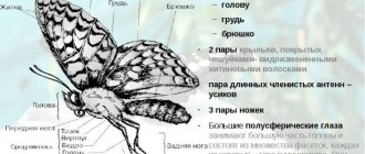
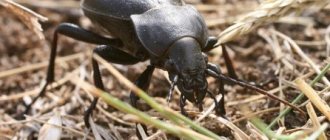
Malarial plasmodium: stages, types, development scheme Malarial plasmodium. Many insects are carriers of dangerous diseases. The malaria mosquito is perhaps the most famous representative of such insects. Malaria is a dangerous disease caused by Plasmodium falciparum. The infection occurs through the bite of a malaria mosquito, the saliva of which may contain specific enzymes and sporozoites of Plasmodium. As a result of a bite, a person experiences severe intoxication of the body, which is accompanied by severe fever. Malaria mosquitoes - carriers of malarial plasmodium. Only some mosquitoes of the genus Anopheles are capable of transmitting malaria. They are called malaria mosquitoes. This is a fairly extensive genus, including several hundred species. Not all representatives of the Anopheles genus are capable of transmitting malaria. Some are very good carriers, while others are not dangerous. These mosquitoes are widespread throughout the world. Including those...
Review
0
Evaluation criterion
Summary: Was the information useful? Please rate it
User rating 4.56 (1 votes)
Malarial plasmodium. Many insects are carriers of dangerous diseases. The malaria mosquito is perhaps the most famous representative of such insects. Malaria is a dangerous disease caused by Plasmodium falciparum. The infection occurs through the bite of a malaria mosquito, the saliva of which may contain specific enzymes and sporozoites of Plasmodium. As a result of a bite, a person experiences severe intoxication of the body, which is accompanied by severe fever.
- 1 Malaria mosquitoes are carriers of malarial plasmodia
- 2 Development cycle of malarial plasmodium
- 3 Periods and symptoms of the disease
- 4 Dimensions of malarial spasmodium 4.1 Sporogony
- 4.2 Tissue schizogony
- 4.3 Erythrocyte schizogony
Malaria mosquitoes - carriers of malarial plasmodium
Only some mosquitoes of the genus Anopheles
. They are called malaria mosquitoes. This is a fairly extensive genus, including several hundred species. Not all representatives of the Anopheles genus are capable of transmitting malaria. Some are very good carriers, while others are not dangerous.
These mosquitoes are widespread throughout the world. Including in those regions where malaria has been eliminated. They are also found in temperate climates; they are also found in the Moscow and Leningrad regions, Siberia, and the Far East.
Therefore, cases of local malaria are also possible in Russia. Since tourists who traveled to disadvantaged regions or migrants can serve as a source of mosquito infection. The risk is especially high in hot years. Mosquito larvae develop in water, so there are many more of them in damp places.
Contrary to popular belief, malaria mosquitoes are small (a few millimeters in length). The easiest way to distinguish them is by their characteristic stance, with the rear end of the body strongly raised upward (other mosquitoes keep their body parallel to the surface).
The presence or absence of the malaria pathogen in the mosquito’s body does not in any way affect its appearance or behavior. Therefore, it is impossible to distinguish between infected and uninfected mosquitoes without special analysis.
After the mosquito has drunk the blood of the patient, time must pass for the malarial plasmodium to go through certain stages of development and the mosquito to become infectious (usually 10–20 days). The rate of development of malarial plasmodia in the body of a mosquito is influenced by many factors, including temperature. At low temperatures, development slows down or stops.
Since the mosquito does not live long, it does not always have time to become infectious, dying before the malaria pathogen completes its development. This is one reason that indigenous malaria is rare in temperate climates. In addition, with the onset of cold weather, the circulation of the pathogen between people and mosquitoes stops.
Mosquitoes themselves do not contain malaria pathogens. To actually become malarial they must drink the blood of a person with malaria. A mosquito can only become infected from a person; malarial plasmodium is not transmitted to its offspring.
One of the main protective measures is to prevent bites from malaria mosquitoes. This is the use of nets and curtains (especially since most malaria mosquitoes prefer to feed at night), spraying insecticides indoors, choosing times and routes, and using repellents.
Plasmodium falciparum is the causative agent of malaria infection. According to the classification of protozoan (single-celled) parasites, they belong to the phylum Apicomplexa, class Sporozoa, order Haemosporida, family Plasmodiidae, genus Plasmodium.
There are 4 types of parasites that can parasitize the human body:
- Plasmodium vivax is the causative agent of tertian malaria;
- Plasmodium malariae is the causative agent of four-day malaria;
- Plasmodium falciparum is the causative agent of tropical malaria;
- Plasmodium ovale is the causative agent of ovale malaria, similar to the species P. vivax.
All varieties are generally similar in shape, structure and life cycles, except for details. But they have their own characteristics of the development cycle; mainly by the duration of its periods.
An interesting feature of parasites of the Apicomplexa type is the ability to alternate sexual (gamogony) and asexual (agamogony) methods of reproduction in the life cycle. In this case, a change of “master” occurs. In the human body, which is an “intermediate host” for the malarial plasmodium, only asexual reproduction is possible. And the phase of sexual reproduction is the prerogative of the main “host” and carrier of malaria, that is, the mosquito.
Skin diseases and sexually transmitted diseases and photos
Dermatology (from the Greek derma - skin and logos - science) is the science that studies skin diseases . Dermatology studies the structure and functions of the skin in normal and pathological conditions, the relationship of skin diseases with various pathological conditions of the body, and also clarifies the causes and pathogenesis of various dermatoses, develops issues of diagnosis, therapy, and prevention of skin diseases. Venereology studies the epidemiology, clinical picture, diagnosis, treatment and prevention of sexually transmitted diseases, which include syphilis, gonorrhea, ureaplasmosis, mycoplasmosis, trichomoniasis, chancroid, chlamydia and other human lesions. For this purpose, clinical, histological, microbiological, immunobiological, functional, biochemical, histochemical, experimental and statistical research methods are used. Dermatology is divided into general and private. The subject of general dermatology is general problems (morphology and physiology of normal and diseased skin, general patterns of development of skin diseases, principles of their treatment and prevention, etc.), and private dermatology is concerned with individual dermatoses and syndromes. Dermatology is closely related to the following disciplines: venereology (syphilis, gonorrhea, ureaplasmosis, mycoplasmosis, trichomoniasis, chlamydia), infectious diseases (herpes, AIDS and HIV infection, exanthema and enanthema), pediatrics (rubella, measles, scarlet fever, chickenpox, etc. ), internal diseases (itching, urticaria, etc.), diseases of the endocrine system (myxedema, etc.), surgical diseases (boils, abscesses, etc.), gynecology and obstetrics (dermatoses of pregnant women), neuropathology (leprosy, syringomyelia, etc. .), ophthalmology, psychiatry, etc. Dermatology is of great importance for practical medicine, since skin diseases account for about 10% of all diseases. Skin diseases are in most cases visible to the naked eye. The characteristics of skin diseases, their availability for research in a variety of ways, and the possibility of direct observation of their evolution favor the study of a number of important physiological problems (immunobiological processes, allergies, reactivity, etc.). In its development, dermatology, which became an independent discipline about 100 years ago, has traveled a difficult path. First, naturally, the clinic of skin diseases was studied, then their histomorphology and microbiology; Recently, pathophysiological, pathogenetic and, finally, biological directions have developed, the foundation of which was the achievements of theoretical medicine. The object of study at present is not only a certain dermatosis, but also the background, the “soil” on which it develops, i.e., the condition of a patient suffering from any skin disease, with all its individual characteristics and living conditions. The medical institutions of our country have been significantly replenished with young doctors who need benefits in dermatology and dermatovenereology, which they need in their daily work. Every practicing physician faces the difficult task of understanding numerous skin diseases. Often, even an experienced dermatologist, when examining a patient, cannot quickly make a diagnosis and is forced to turn to reference literature to substantiate his conclusion. It is quite understandable that inexperienced doctors experience much greater difficulties with this. In our opinion, every practicing doctor should always have with him a reference book on dermatology, in which he can find a description of a suspected skin disease, prescribe rational treatment and outline a plan of preventive measures. Considering the wider distribution of certain forms of skin diseases, we considered it appropriate to dwell on these diseases in a little more detail. Particular attention is paid to pyoderma, which is often found among industrial and agricultural workers. This section highlights the classification of pyoderma, describes various clinical forms, their prevention and treatment. In addition to well-known skin diseases, the reference material contains a large number of syndromes, symptom complexes, as well as individual skin diseases , designated by the name of the author. It includes only those syndromes and diseases that have well-defined clinical symptoms and have received general recognition in dermatology. Among infectious diseases, a special place is occupied by skin tuberculosis and leprosy; in this regard, the site outlines the clinical variants of these diseases, their epidemiology and modern methods of therapy. Compared to skin diseases, sexually transmitted diseases (syphilis, gonorrhea, ureaplasmosis, mycoplasmosis, trichomoniasis, chlamydia), which are encountered by doctors of many clinical disciplines, are described in somewhat more detail than skin diseases. To successfully combat these sexually transmitted diseases, doctors are obliged not only to make a correct diagnosis in a timely manner, but also to take an active part in preventive measures. A lot of time has passed since work on the site began. During this time, dermatology received new treatment methods (photochemotherapy, etc.). The number of drugs used in the treatment of skin diseases, various syndromes and symptom complexes has increased. Some drugs have been discontinued. This necessitated a number of corrections and additions. In the proposed material, the sections “Gonorrhea”, “Syphilis”, “Ureaplasmosis”, “Trichomoniasis”, “Mycoplasmosis”, “Chlamydia”, “Herpes”, “AIDS”, “HIV infection”, “Rubella”, “ Measles", "Scarlet fever", "Chickenpox". Skin and venereal diseases are very diverse in their clinical pathology and occur quite often. Therefore, doctors of all clinical specialties often have to deal with patients with skin and venereal diseases. In addition, skin changes often serve only as an external reflection of one or another pathology of internal organs, the central nervous system, or severe systemic diseases. Dermatology and venereology is by no means a “narrow” specialty that only a few “true” dermatologists should master. On the contrary, knowledge of the basics and elements of dermatology and venereology is necessary in the daily work of a doctor of any specialty, including a dentist. The ability to understand these issues should be considered one of the mandatory aspects of professional training, the official duty of a doctor in any clinical specialty. The specifics of teaching dermatovenereology at the Faculty of Dentistry include direct medical guidance, manipulation, prevention and medical examination of patients with diseases of the oral mucosa, which very often are symptoms of many skin diseases and syphilis. Therefore, the main attention should be paid to the study of changes in the oral mucosa during skin and venereal diseases, their clinical varieties, pathogenesis and special treatment. Particular attention should be paid to skin diseases, which often, and sometimes in isolation, appear on the mucous membrane of the mouth and lips. This applies to lichen planus, blistering diseases, exudative erythema multiforme, lupus erythematosus, cheilitis, candidiasis, herpes, etc. A number of diseases in which the mucous membrane of the mouth and lips are affected relatively rarely, for example tuberculosis, leprosy, pustular psoriasis, etc. are offered to future dentists in order to create their understanding of these diseases, without deepening and detailing the symptoms of the disease and treatment. The future dentist must also have an understanding of common skin diseases in order to provide advisory or emergency assistance. These are pyoderma, dermatitis and toxiderma, urticaria, etc. The existing textbooks on dermatovenerology are not sufficiently oriented for teaching skin and venereal diseases at dental faculties and do not fully correspond to the goals and objectives of the training of dentists provided for by the curriculum on skin and venereal diseases. With many skin diseases and syphilis, quite often rashes appear not only on the skin, but simultaneously in the oral cavity, and often skin diseases begin with damage to the oral mucosa, and skin rashes appear much later or the disease manifests itself only as rashes in the mouth or on the red border lips In the latter cases, the main role in diagnosing skin diseases, and sometimes in their treatment and prevention of relapses belongs to dentists. Dermatology as a science was defined in the 18th century. Existed since the 19th century. German, English, French and Russian schools of dermatology at first differed sharply in matters of understanding the essence of skin diseases, but by the beginning of the 20th century. these differences have been smoothed out, and now we can only talk about some features of the direction of scientific research of one or another school, and not about different understandings of the etiology and pathogenesis of diseases. International congresses of dermatologists, dermatological journals, etc. played a huge role in this issue. It should be emphasized that for domestic dermatology, the founders of which were A. G. Polotebnov (1838-1907), A. I. Pospelov (1846-1916), etc., there has always been Characteristic is the attitude towards the patient from the perspective of the whole organism, highlighting the leading role of the nervous system in the pathogenesis of many skin diseases. In the study of the pathology of the oral mucosa, including skin and venereal diseases, the main role belongs to dermatologists, who have described the vast majority of diseases in this area. For example, Setton described deep recurrent aphthae, Turkish dermatologist Behçet - a disease later named after him, Wilson - damage to the oral mucosa with lichen planus, A. I. Pospelov - with psoriasis, Dubrnel - lupus erythematosus on the oral mucosa, Gebra – exudative erythema multiforme, etc. A great contribution was made by Soviet dermatologists, in particular employees of the department of skin and venereal diseases of the Moscow Medical Dental Institute. N. A Semashko. The founder of the dermatostomatological direction was the first head of the department B. M. Pashkov (1899–1973). He belongs to the first description of soft leukoplakia, together with P. D. Sheklakov - benign non-acantholytic pemphigus of the oral mucosa only, the identification of two clinical forms of exfoliative cheilitis - dry and exudative, etc. After B. M. Pashkov, his student A. L became the head of the department Mashkilleyson, who described new forms of precancerous diseases of the lips, meteorological cheilitis, together with T. N. Antonova and E. I. Abramova - vesical-vascular syndrome of the oral mucosa and other diseases. Employees of the department have studied in detail various clinical forms of lichen planus on the oral mucosa and lips, showing the role of gastrointestinal pathology in the pathogenesis of this disease, as well as leukoplakia; various clinical forms of lupus erythematosus on the oral mucosa and red border of the lips are described; classifications of precancerous diseases of the mucous membrane of the mouth and lips, cheilitis have been developed; atopic cheilitis is isolated; the pathogenesis has been studied and treatment for exfoliative cheilitis, Melkersson–Rosenthal syndrome, exudative erythema multiforme has been developed; cystic dermatoses, psoriasis, Behçet's disease, furunculosis, etc. Skin and venereal diseases of adults and children have their own specifics. Due to the significant difference in the reactivity of the body of an adult and a child, in the endocrine status, in the state of the central nervous system, as well as due to the intensive growth of the child’s body and other factors, the same skin diseases often clinically occur with significant differences. In addition, some skin diseases are characteristic mainly or only of adulthood or childhood. For example, occupational skin diseases occur only in adults, epidemic pemphigus, pseudofurunculosis and exfoliative dermatitis - in newborns; Early congenital syphilis manifests itself before the age of 4 years, and late congenital syphilis occurs in the vast majority of cases between 7 and 16–18 years of age. The course of acquired syphilis in children has its own characteristics. These circumstances prompted the creation of separate programs for teaching skin and venereal diseases in the medical and pediatric faculties of medical institutes and textbooks on skin and venereal diseases intended for students of either medical or pediatric faculties. However, a significant number of skin diseases occur in the same way or with slight differences in both adults and children. In addition, practice shows that graduates of the Faculty of Medicine often have to admit children with skin diseases, diagnose and treat them, and graduates of the Faculty of Pediatrics have to do this for adults. Particular attention is paid to protecting the health of mothers and children, steadily improving the quality of medical care, expanding clinical examination, developing new methods and means of prevention, diagnosis and treatment of the most common skin diseases, including dermatoses in children. Skin diseases are an important medical, biological and social problem that requires urgent solutions. It has been established (I.V. Shchutsky) that in recent years the incidence of skin diseases among the child population is 11-12%, and allergic dermatoses are observed in 32% of children under three years of age. 80-95% of children suffering from chronic skin diseases are diagnosed with diseases of the liver, pancreas, and bile ducts, which require special treatment in a hospital setting. Similar results were obtained by N.P. Toropova and co-authors. Skin diseases in children, as a rule, occur against the background of severe pathology of internal organs. Their treatment on an outpatient basis is associated with the release of most mothers from socially useful work in caring for children and with a significant loss of labor resources. The child’s body at different periods of life is distinguished by characteristic anatomical and physiological features, allergic and immunological reactivity. This is due to the influence of hereditary, constitutional, metabolic, neuroendocrine regulatory mechanisms on the formation of the child’s body, including the skin and its functions. The development of skin pathology at different age periods is also significantly influenced by environmental, social and everyday factors, regimen, nutrition, and hygienic care of the child’s skin. Unlike adults, there are some features of the treatment of skin diseases during different periods of childhood. As a result of the constant concern of the Soviet state for the health of mothers and children in our country, in recent years, dermatological offices have been opened at children's clinics, and the network of dermatological departments at children's hospitals and dermatovenerological dispensaries has been expanded. The country's medical institutions have been replenished with young doctors who feel the need for manuals and reference books on pediatric dermatovenerology, which they need in their daily work. The site provides the basic principles of general and local treatment of skin diseases in children. The description of dermatosis ends with recommendations for prevention and clinical monitoring of the child in order to prevent relapses of the disease. At the end of each disease, doses and methods of use of medications most commonly used in pediatric dermatology are given. When performing such labor-intensive and multifaceted work, it was impossible to avoid some shortcomings. In this regard, comments and suggestions from readers will help improve the quality of the material presented in further work on it and will be received with gratitude.
Developmental cycle of malarial plasmodium
Malarial plasmodium. The development cycle of malarial plasmodium is quite complex. When a mosquito infected with malaria bites, sporozoites enter the human bloodstream (see below). In the human body, sporozoites primarily enter the cells of the reticuloendothelial system, forming primary tissue forms there.
This period of development of malarial plasmodium is asymptomatic. Subsequently, malarial plasmodia enter the blood and penetrate into red blood cells, completing a cycle of asexual development and reproduction there (schizogony).
The duration of one cycle of schizogony depends on the type of malarial plasmodium and determines the cyclicity of febrile attacks. Asexual forms of Plasmodium falciparum are called schizonts.
A mature schizont is divided into merozoites in the erythrocyte. After this, the erythrocyte is destroyed, and merozoites are released into the blood and then penetrate into new erythrocytes, undergoing a new development cycle there.
Find out more: Klebsiella in stool in adults: symptoms and treatment
During tertian malaria, some of the merozoites can enter the cells of the reticuloendothelial system and form secondary tissue forms. The presence of secondary tissue forms determines the possibility of relapses of malaria.
Along with asexual forms (schizonts), sex cells can also be formed in erythrocytes - gametes: male (microgametes) and female (macrogametes). The presence of sexual forms of plasmodium in the blood does not give clinical manifestations, but is dangerous from an epidemiological point of view: such patients are infectious to mosquitoes.
Gametes, entering the mosquito’s body, undergo a sexual development cycle there, leading to the formation of a huge number of sporozoites that penetrate the salivary glands of the mosquito and are capable of infecting humans.
Life cycle of Plasmodium falciparum in humans: erythrocyte (clinical) stage of malaria
After the liver cells rupture, merozoites enter the blood and invade red blood cells. The erythrocyte (clinical) stage of schizogony begins.
Rice. 14. Erythrocyte schizogony. 5 and 6 are ring-shaped trophozoites. 7, 8 and 9 - young, immature and mature schizonts. 10 - erythrocyte merozoites.
Attachment to red blood cells
Attachment of merozoites to the membrane of erythrocytes and invagination into their membranes occurs due to the presence of special receptors on the surface of red blood cells. It is believed that the receptors on the surface of erythrocytes that serve as targets for merozoites are different for different types of Plasmodium.
Rice. 15. Red blood cells infected with Plasmodium vivax (the causative agent of tertian malaria) and Plasmodium ovale (the causative agent of tertian malaria) become enlarged, discolored and deformed, and toxic granularity appears in them. When infected with Plasmodium malariae (the causative agents of four-day malaria) and Plasmodium falciparum (the causative agents of tropical malaria), the shape and size of red blood cells do not change.
Erythrocyte schizogony
Having penetrated red blood cells, schizonts absorb the protein globin (a component of hemoglobin), grow and multiply.
In the erythrocyte, the parasite goes through 4 stages of development:
- Ring (trophozoid) stage.
- Stage of amoeboid schizont.
- Morula (fragmentation) stage. At this stage, the nuclei of schizonts are repeatedly divided (into 6 - 25 parts), and areas of the cytoplasm are separated around them. Erythrocyte merozoites are formed.
- Some parasites go through the stage of gametocyte formation.
A parasite that has penetrated an erythrocyte is called a trophozoid (schizont). It gradually increases in size. A vacuole appears near its nucleus and the trophozoid takes the form of a ring (ring) - a ring-shaped schizont.
As they grow (schizonts feed on hemoglobin), they increase in size and take on the appearance of an amoeba - an amoeba-shaped schizont.
Then, as the schizont grows, it becomes rounded and its nucleus divides many times—the morula stage. Each type of plasmodia has a certain number of nuclei: 12 - 12 in P. vivax, 6 - 12 in P. malariae and P. ovale, 12 - 24 in P. falciparum. This is how erythrocyte merozoites are formed. From the 1st schizont, 8–24 blood merozoites are formed, from which asexual and sexual forms of parasites further develop.
The duration of the erythrocyte schizogony phase is 72 hours in P. malariae, and 48 hours in other Plasmodium species.
During growth, pigment accumulates in the cytoplasm of parasites. Its appearance is associated with the processes of hemoglobin assimilation. Hemoglobin accumulations look like rods or grains. Its color ranges from golden yellow to dark brown.
Rice. 16. During growth, pigment accumulates in the cytoplasm of parasites.
After the destruction of erythrocytes, merozoites enter the blood, some of which re-enter the erythrocytes, others undergo a cycle of gametogony - transformation into immature germ cells, gamonts.
Together with merozoites, heme (the second component of hemoglobin) enters the blood. Heme is a powerful poison and causes acute attacks of malarial fever.
Cycles of erythrocyte schizogony are repeated every 3 days, in other types of malarial plasmodia - every 2 days.
Rice. 17. Destruction of the erythrocyte and release of merozoites into the blood.
Rice. 18. A merozoite of Plasmodium vivax (the causative agent of tertian malaria) that has penetrated into an erythrocyte (thin blood smear).
Rice. 19. Young trophozoids of Plasmodium falciparum (the causative agent of tropical malaria) under a microscope.
Rice. 20. Ring-shaped trophozoites of Plasmodium vivax, the causative agents of tertian malaria (ring stage).
Rice. 21. The photo shows an amoeboid schizont Plasmodium vivax (stage of an amoeboid schizont).
Rice. 22. The photo shows mature Plasmodium vivax schizonts (morula or fragmentation stage).
When red blood cells are destroyed and merozoites are released into the plasma, febrile attacks and anemia develop. When liver cells are destroyed, hepatitis develops.
Periods and symptoms of the disease
Malarial plasmodium. The entire process, from infection to recovery, can be divided into four periods: incubation, acute, latent and relapse. The nature of the course of the disease and the frequency of relapses largely depend on the type of malarial plasmodium - the causative agent. For example, the incubation period of the disease can vary from 7 days to 1 year, depending on the type of malaria infection.
General characteristic signs of the disease:
- elevated body temperature (39 – 40 degrees); chills and fever replace each other;
- “ache” throughout the body;
- prolonged headaches;
- gagging and diarrhea;
- profuse sweating after a hot flash;
- severe enlargement of the liver and spleen;
- pain in the liver area;
- general weakness and anemia;
- possible confusion;
- yellowing of the skin and sclera of the eyes;
- frequent and painful urination;
- loss of appetite;
- insomnia.
Individual signs and symptoms (for example: cardiac stabbing pain, asthma attacks, joint pain) are also possible and depend on the presence of “weak points” in the body and the functioning of the immune system. Sick children usually develop a rash on their body. Tremors of the limbs are also typical for infants.
As the underlying disease progresses, other pathologies may occur, such as cerebral vascular ischemia and necrobiosis in the kidneys. In case of complications of the disease, coma and rupture of the spleen are possible.
Sizes of malarial spasmodium
The life cycle of human malaria pathogens consists of the following stages: the sexual process of reproduction in mosquitoes (sporogony); asexual reproduction in liver cells (tissue schizogony); asexual reproduction in erythrocytes (erythrocyte schizogony); formation of sexual forms in erythrocytes - gametocytes.
Sporogony
In the body of a nopheles patient with malaria or a parasite carrier, gametocytes enter the stomach of the insect: macrogametocytes (female) and microgametocytes (male). After restructuring of the nuclear apparatus, the macrogametocyte turns into a macrogamete. 4-8 microgametes are formed from a microgametocyte.
In the stomach of a mosquito, fertilization of a macrogamete by a microgamete occurs. As a result, a motile zygote is formed, called an ookinete. The latter penetrates the wall of the stomach and an oocyst forms on its outer side.
The nuclei in the oocyst divide repeatedly, after which sporozoites are formed inside the oocyst - elongated spindle-shaped bodies measuring 11-15 microns in length.
The oocyst shell ruptures and the sporozoites penetrate the salivary glands. When a mosquito bites, sporozoites enter the human body. The duration of sporogony depends on temperature. At temperatures below 16°, sporogony does not occur.
Tissue schizogony
Malarial plasmodium. Sporozoites can remain in human blood for no more than one hour. During this period, they penetrate the liver parenchyma cells, where schizonts are formed. Their development has been studied in detail on Plasmodium species parasitizing monkeys and partly on human Plasmodium.
The sporozoite, having penetrated the liver cell, becomes rounded, increases in size, and the nucleus of the resulting schizont is successively divided. By the 6-12th day, the parasite fills the entire liver cell, pushing the cell nucleus to the periphery.
The size of the parasite is up to 60 microns. Such a large schizont breaks up into a large number (thousands and tens of thousands) of merozoites.
These latter in P. falciparum penetrate erythrocytes and subsequently the parasites develop only in erythrocytes; in other species, merozoites penetrate erythrocytes, as well as into liver parenchyma cells, where they undergo subsequent cycles of tissue schizogony. The duration of tissue development in P. vivax vivax and P. ovale is 7-8 days, P. malariae 11-12 days.
Erythrocyte schizogony
Tissue merozoites penetrate the erythrocyte and form schizonts, which disintegrate into erythrocyte merozoites. Red blood cells are destroyed and the released merozoites settle in new red blood cells.
Find out more: Ureaplasmosis - causes, signs, symptoms and treatment
Malarial plasmodium. And some of the merozoites form gametocytes. The latter can circulate in the blood for a long time, their further development (sporogony) occurs in the carrier. Different stages of development of malaria pathogens in the blood are clearly distinguishable by morphological characteristics. However, P. vivax vivax cannot be distinguished from P. vivax hibernans.
The morphological characteristics of the pathogens themselves and the changes they cause in red blood cells make it possible to determine the type of parasite using preparations (smears and thick drops of blood). In the erythrocyte development cycle of Plasmodium, the following stages are distinguished: rings, schizont, merulation, young and formed gametocytes.
The size and shape of the parasite at different stages of development, the duration of the entire schizogony cycle, the number of merozoites formed and their size, the number of grains (lumps) of pigment, their color and location in the parasite, the shape, size and other characteristics of gametocytes, as well as changes observed in the infested erythrocytes, serve as signs to distinguish one species from another.
The development of the erythrocyte stages of all types of human malaria pathogens occurs in the circulating blood. An exception is P. falciparum, in which only ring stages and gametocytes are found in the blood; further development of schizonts up to the release of merozoites from erythrocytes occurs in the capillaries in which the deposited blood is located.
Infectious diseases.
Infectious diseases have occupied a major place in human pathology for many centuries and have been a scourge for the population. In pre-revolutionary Russia, millions of people fell ill with various infectious diseases every year; there was no year free from epidemics. Infectious diseases were the main cause of child mortality and general mortality of the population; they caused enormous damage to the population and the country's economy because they often left severe and irreversible consequences. Tens of thousands were blind after smallpox, deaf, deaf-mute after scarlet fever, and had persistent paresis and paralysis after meningitis and polio. The Soviet state faced a difficult situation with regard to infectious diseases from the very first days of its formation. It was further aggravated by the previous world war, the civil war that followed the revolution, foreign intervention, famine and devastation, and the huge migration of the population in the country. All this taken together led to the emergence of epidemics of unprecedented scale. Already in those days, doctors attached great importance to the fight against infectious diseases. Already among the first acts of the Soviet state were decrees and resolutions to combat these diseases. At the beginning of the 20th century, the fight against infectious diseases unfolded on a broad front. First of all, appropriate personnel were required. This problem was solved thanks to the organization of appropriate departments at medical institutes. No less importance was attached to the study of infectious diseases, which included specialists from various fields. Gradually, corresponding laboratories and institutes were created, in which (together with the departments of medical institutes) issues of infectious pathology were developed. Children's infectious diseases were isolated in a separate course because the most common infections, more often or almost exclusively, occur in childhood and even mainly in early or preschool age (they were even considered obligatory for children). For example, the incidence of measles was almost universal among children and was observed more often under the age of 5 years. Among those sick with whooping cough (the second most common childhood infection), half were children under 3 years of age. The incidence of chickenpox and scarlet fever is highest between the ages of 1 and 9–10 years. Infectious diseases took first place in the structure of child mortality, which was very high. Finally, all infectious diseases, including those that are common among both children and adults, such as dysentery, in children have a number of pathogenetic and clinical features due to the anatomical and physiological differences of the child’s body. In the 1920s, a consistently expanding study of infectious diseases in children began. It is necessary to note the leading importance of the work carried out by teams of scientists led by A. I. Dobrokhotova, M. G. Danilevich, A. A. Koltypin, V. I. Molchanov, and subsequently their students and followers. An exceptionally large contribution to the study of the pathogenesis of infectious diseases was made by pathomorphologists (V.D. Tsinzerling, A.A. Skvortsov, etc.). Various studies were carried out by domestic immunologists, microbiologists, virologists and other specialists; The most active work was carried out by epidemiologists. A huge amount of research has been carried out, unique literature has been created, and a coherent system for combating childhood infectious diseases has been developed, as a result of which unprecedented successes have been achieved in history. In our country, the incidence of morbidity has sharply decreased - even to the point of eliminating certain nosological forms; for all infectious diseases, the mortality rate has decreased, the mortality rate has decreased hundreds and tens of times; Childhood infections have almost lost their significance in population mortality. The sharply increased life expectancy in our country is generally recognized as the result of a decrease in child mortality and mainly due to childhood infections. A coherent system of combating infectious diseases has a state character. The work of healthcare workers, in particular their fight against infectious diseases, is an important part of the general creative activity of modern society. This fight is not over; the tasks of further neutralizing childhood infections remain to be solved. Currently, intensive development in the field of infectious diseases continues and leads to the discovery of new aspects of known diseases, the identification of new pathogens and even new nosological forms. Infectious diseases, under the influence of numerous factors, have undergone evolution, especially noticeable in recent decades. Therefore, the presentation of the course is characterized by increasing complexity and versatility when considering each nosological unit. A description of the classic manifestations of infections, characteristics of new forms of infectious diseases that have appeared in the process of evolution, the reason for their formation and, finally, modern features of pathogenesis and clinic are presented. Infectious diseases are diseases caused by microorganisms. They are distinguished by their contagiousness, the specificity of the pathogen, clinical patterns in the form of cyclicity and the formation of immunity during the disease process. Contagiousness consists in the transmission of the pathogen from the patient to surrounding people. The specificity of the pathogen is that this microorganism, when infecting other people, causes the same disease in them and this nosological form cannot be caused by other microorganisms. For example, typhoid fever, dysentery, diphtheria, and measles are caused, respectively, by typhoid bacilli, dysentery, and measles virus. Some other microorganisms may cause similar individual symptoms, syndromes, but not the entire complex of the disease. An infectious disease is often referred to as “infection.” However, these are not synonyms; the concept of “infection” is much broader and, along with infectious diseases, includes the so-called infectious inflammatory processes. The latter are also caused by microorganisms and are contagious, but do not have pronounced clinical patterns in the form of cyclicity divided into certain periods. The main difference is that the pathogen does not cause any specific clinical form, but a variety of diseases with their own clinical picture. Similar, up to complete clinical identity, diseases can be caused by other types of pathogens, for example, streptococcal, staphylococcal processes. Both streptococci and staphylococci have the ability to infect any tissue, any organ of the human body and cause various diseases: rhinitis, tonsillitis, otitis media, lymphadenitis, pneumonia, pleurisy, meningitis, sepsis, etc. Many of these diseases can be caused by other microorganisms. Thus, pneumonia can be caused not only by streptococci and staphylococci, but also by Pfeiffer’s bacillus, Klebsiella, etc. These infectious processes have not taken shape in the form of a specific infectious disease with its inherent cyclicity, their nosological characteristics are not sufficiently delineated. They usually occur in the form of inflammatory processes, their infectious nature is immutable. There are also differences regarding immunity. Infectious diseases provide predominantly strong immunity not only for years, but often for life (measles, chicken pox, mumps, whooping cough, typhoid fever, scarlet fever, etc.). Infectious processes either do not provide immunity at all or it is short-lived; in some cases there is even a tendency to recurrence of diseases, as is observed in relation to streptococcal and staphylococcal diseases. The division between infectious diseases and infectious processes is often very arbitrary; as we study, the lines between them become noticeably blurred. Individual infectious processes are united on the basis of common clinical, morphological and other signs, and most importantly - on the basis of the unity of the pathogen. Thus, streptococcal and staphylococcal infections were isolated and included in textbooks on infectious diseases. The main differences between infectious diseases are contagiousness, the specificity of the pathogen, the peculiarity of clinical patterns, and the formation of immunity. During infectious diseases, there is an incubation, or latent, period, a prodromal, or period of precursors, periods of disease development and convalescence. Infectious pathology must be understood in a broad sense, including all diseases caused by microorganisms - both infectious diseases and infectious processes.
The structure of malarial plasmodium
Malarial plasmodium. Phylum Sporozoa: the phylum includes exclusively parasitic protozoa. In connection with the parasitic lifestyle, a simplification of the organization occurs (the disappearance of organelles for capturing and ingesting food, digestive and contractile vacuoles). The life cycle becomes more complex - a change of hosts, alternating asexual and sexual reproduction. The representative of the phylum is the malarial plasmodium.
Sporozoites are thin, worm-shaped cells that enter the liver cells through the bloodstream, where they transform into schizonts, which reproduce by multiple divisions - schizogony. In this case, the nucleus divides repeatedly, then a large number of daughter cells are formed from each cell.
The resulting merozoites leave the liver cells and invade the red blood cells. Here they feed, then schizogony occurs again. Thus, two forms of schizogony are distinguished - in liver cells and in erythrocytes.
Plasmodium falciparum causes malaria in humans. Infection occurs through the bite of a malarial mosquito (genus Anopheles), which contains the pathogen at the sporozoite stage.
As a result of erythrocyte schizogony, 10-20 merozoites are formed, which destroy the erythrocyte, enter the blood and infect subsequent erythrocytes. The cyclical nature of malaria attacks is due to the cyclical release of merozoites and their metabolic products from erythrocytes into the blood plasma.
After several cycles of schizogony, gamonts are formed in erythrocytes, which in the mosquito’s body will turn into macrogametes and microgametes. When gamonts enter the stomach of a mosquito, they turn into gametes, copulation occurs, the fusion of gametes. The zygote is mobile and is called an ookinete.
The ookinete migrates through the mosquito's stomach wall and develops into an oocyst. The oocyst nucleus divides many times, and the oocyst breaks up into a huge number of sporozoites - up to 10,000. This process is called sporogony. Sporozoites migrate to the salivary glands of the mosquito. Meiosis occurs after the formation of the zygote, sporozoites are haploid.
Thus, in the life cycle of Plasmodium falciparum, humans are the intermediate host (pre-erythrocytic schizogony, erythrocyte schizogony, onset of gametogony), and the malarial mosquito is the final host (completion of gametogony, fertilization and sporogony).
Life cycle of Plasmodium falciparum in humans: exoerythrocytic (preclinical) stage of malaria
Infection
When an infected female mosquito bites a person, malarial plasmodia at the sporozoite stage enter the human bloodstream with the saliva of the insect. Within 10 to 30 minutes, sporozoites move freely in the blood plasma and then settle in the liver cells. Some of the sporozoites (bradysporozoites) Plasmodium ovale and Plasmodium vivax hibernate, while another part of them, as well as Plasmodium falciparum and Plasmodium malariae (tachysporozoites) begin hepatic schizogony immediately.
Rice. 12. Tissue exoerythrocytic schizogony. 2 - trophozoite, 3 - schizont, 4 - release of merozoites from liver cells into the blood.
Period of tissue schizogony
In liver cells (hepatocytes), the transformation of sporozoites into tissue schizonts occurs, which after 6–15 days divide to form many tissue merozoites. From one sporozoite, from 10 to 50 thousand hepatic merozoites (schizonts) are formed, which enter the blood after 1 - 6 weeks.
When infected liver cells are destroyed, tissue merozoites are released into the blood. This ends the incubation period of malaria and begins the period of erythrocyte schizogony - the period of clinical manifestations.
Hibernation process
Some sporozoites (hypnozoites) of Plasmodium ovale and Plasmodium vivax, once in hepatocytes, turn into inactive forms and hibernate. Parasites can remain in this state for months and years, causing distant relapses.
Rice. 13. Tissue schizont in the liver.
Schizogony
Malarial plasmodium. Plasmodium vivax causes three-day malaria, Plasmodium malariae - four-day malaria, Plasmodium falciparum - tropical, Plasmodium ovale - oval malaria, which is mainly characteristic of the inhabitants of Central Africa.
The French physiologist Charles Louis Alphonse Laveran discovered the human malarial plasmodium in 1890. Since 2004, another species has been identified - Plasmodium knowlesi, living in Southeast Asia. In addition, the latter type can cause malaria in long-tailed macaques (cynomolgus macaques, or cynomolgus macaques).
When bitten by the Anopheles mosquito, saliva enters the human bloodstream, containing specific enzymes that block blood clotting and Plasmodium sporozoites. Sporozoites are curved, fusiform, up to 15 µm long forms of plasmodium.
Find out more: Amoeba vulgaris: structure, respiration, nutrition
With the blood flow through the vessels, the sporozoites enter the human liver cells, where the asexual reproduction of the parasite begins. Schizogony of plasmodiums has distinctive features: the mother cell as a result of division produces not 2 daughter cells, as in other representatives, but many.
During tissue and erythrocyte schizogony, the formation of digestive vacuoles occurs in the protoplasm (internal contents of the cell) of merozoites. In these vacuoles, the nutrients necessary for plasmodium accumulate and waste products (toxins) that are not needed by plasmodium and are harmful to humans are removed from them.
Not all erythrocytes form new merozoites; in some, the formation of male and female germ cells - gamonts (hemotocytes) - occurs. Coming out of destroyed erythrocytes, merozoites penetrate into other (healthy) ones and divide again, soon destroying these erythrocytes as well.
Such repeated transitions occur with a constant frequency: in Plasmodium malariae every 72 hours, and in other species of Plasmodium - every 48 hours. With the same frequency, a patient with malaria experiences symptoms of intoxication (as toxins are released into the blood): chills and very high body temperature. These cycles of erythrocyte schizogony will be repeated until the number of merozoites necessary for further development is formed.
During tissue and erythrocyte schizogony, the formation of digestive vacuoles occurs in the protoplasm (internal contents of the cell) of merozoites. In these vacuoles, the nutrients necessary for plasmodium accumulate and waste products (toxins) that are not needed by plasmodium and harmful to humans are removed from them. Not all erythrocytes form new merozoites; in some, the formation of male and female germ cells - gamonts (hemotocytes) - occurs.
Tropical diseases.
The site includes basic information about the etiology, pathogenesis, clinical picture, diagnosis, treatment and prevention of protozoal, bacterial, viral and fungal diseases common in hot countries. In addition, data is presented on the hygiene of hot countries and anti-epidemic measures to combat infectious diseases in countries with a tropical climate. Teaching and personal experience in medical institutions in some countries of Africa and Asia were the basis for the creation of this textbook on the main sections of the course of tropical medicine. Tropical diseases are based on sections of the official program for foreign students studying at medical institutes and universities of the CIS countries; the main sections of the course of tropical medicine are considered here. When writing the textbook, the authors did not pretend to be an exhaustive presentation of all nosological forms of diseases found in countries with tropical and subtropical climate, but they meant. The site covers selected issues of tropical medicine, which included protozoal and helminthic infestations, viral infections, and tropical mycoses. The proposed material reflects particularly dangerous infections (smallpox, cholera, plague) and diseases caused by poisonous animals. The greatest share is occupied by clinical issues, diagnosis and treatment of the actual tropical forms of parasitic and vector-borne diseases and mycosis. When creating this section of the site, we used the data accumulated to date in domestic and foreign literature, as well as personal experience in epidemiology, clinical presentation, diagnosis, and treatment of diseases characteristic of subtropical and tropical climate zones. One of the important issues, the presentation of which requires unification, is the nomenclature of chemotherapeutic drugs.
Medical news in the world:
| Is coffee harmful or beneficial? Regular coffee drinkers do not experience an increase in blood pressure after caffeine administration. In irregular coffee drinkers, blood pressure increases after taking both caffeine and coffee or CBC; therefore, it can be assumed that other ingredients in coffee, rather than caffeine, are responsible for this increase in BP. Read more… | Pregnancy – no AIDS! Scientists have concluded that highly active antiretroviral therapy (HART) significantly reduces the risk of transmitting HIV infection to children (transmission of infection to children from mothers who received ART was observed in only 1% of cases), and also helps today to lead a normal, comfortable lifestyle . Read more… | Raw milk can cause serious pregnancy complications A chemical compound found in unpasteurized food was found at unusually high levels in the red blood cells of pregnant women diagnosed with preeclampsia, a serious pregnancy complication that can lead to heart problems. Read more… | Fish oil can be harmful Fish oil supplements that help some heart patients may worsen the condition of others, reports EurekAlert, citing a study from St. Michael and the University of Toronto, Canada, published in yesterday's issue of the Journal of the Canadian Medical Association. Read more… |
| Treatment for impotence has been found!!! Recently, scientists managed to isolate a substance that enhances erection, but acts differently than Viagra (and similar drugs) from spider venom. Read more… | New methods of dental treatment. Toothache let's say: “NO!” ...the discovery of this gene will help recreate damaged tooth enamel, facilitate dental treatment, and even grow new teeth to replace lost ones. Read more… | Curing cancer in 60 minutes is a reality. CyberKnife works with submillimeter resolution, allowing tumors to be clearly localized and eliminated so that the loss of surrounding healthy tissue is minimal - up to 0.1–0.3 mm. Read more… | Karakurt or Black Widow is already in our area. The Black Widow's venom is stronger than that of one of the most dangerous snakes - the rattlesnake (15 times stronger). Read more… |
Sporogony
After several cycles of schizogony have occurred and gamonts have formed, the next stage of the plasmodium life cycle will begin. But for this it is necessary to make the transition into the body of the main host - the mosquito. This occurs when a mosquito bites an infected person. Together with the blood that drinks, gamonts enter the mosquito’s body.
Then, in the cavity of the mosquito’s stomach, the formation of mature germ cells - gametes - from gamonts and their fusion (fertilization) occurs. As a result of fertilization, a zygote is formed that penetrates the wall of the stomach.
Mature sporozoites from the wall of the mosquito's stomach pass into its salivary glands, and then, when a mosquito bites a person, they enter the human blood with saliva. And from this moment the entire life cycle of the malarial plasmodium begins anew.
Here the zygote becomes a growing and developing oocyst, which divides repeatedly to form thousands of new sporozoites. The process of formation of new sporozoites capable of reproduction through schizogony in the mosquito body lasts 7-45 days. This duration is influenced by the ambient temperature: the higher it is, the faster the sporogony stage.