Malaria is an insidious disease that kills millions of people every year. Despite the existence of effective medications, treating the disease is not an easy task. The fact is that the causative agent of the infection is the parasitic microorganism Plasmodium malaria. Its development cycle is quite complex. For this reason, the disease can occur in a latent, chronic or acute form, and also have relapses.
In our publication we would like to talk about who is the intermediate and main host of the malarial plasmodium. We will also consider the reproduction features of the pathogen and its life cycle.
Malarial plasmodium: characteristics
The presented infectious agent belongs to the category of protozoan microorganisms of the genus Plasmodium. There are thousands of single-celled creatures in the world that belong to this genus. However, malaria is caused by only a few species, which are found in tropical areas of Asia and Africa.
All Plasmodium falciparum species are eukaryotes. In other words, the basis of their single-celled organism is the nucleus, in which all genetic information is stored. A specific feature is that the reproduction of malarial plasmodium occurs in human red blood cells. The parasite is characterized by all sorts of transformations, which can affect the state of the body of the carrier of the disease in the most unexpected way.
The life cycle of Plasmodium falciparum is quite complex. It often has a long period. This is due to the need of the parasite to change its development environment. We are talking about the movement of a microscopic infection in separate forms from the main to the intermediate host and back.
Recommendations
- "CDC - Malaria Parasites - About Us." CDC: Malaria
. US Centers for Disease Control and Prevention. Retrieved December 28, 2015. - ^ a b
Zilversmith, M.;
Perkins, S. " Plasmodium
." Web project “Tree of Life”. Retrieved June 1, 2016. - Obado, Samson O; Glover, Lucy; Deitch, Kirk W. (2016). "Nuclear envelope and gene organization in parasitic protozoa: disease-associated specializations". Molecular and Biochemical Parasitology
.
209
(1–2): 104–113. Doi:10.1016/j.molbiopara.2016.07.008. PMID 27475118. - ^ a b
Jimenez-Ruiz, Elena;
Morlon-Guillot, Juliette; Daher, Wassim; Meissner, Markus (2016). "Mechanisms of vacuolar protein sorting in apicomplexan parasites". Molecular and Biochemical Parasitology
.
209
(1–2): 18–25. Doi:10.1016/j.molbiopara.2016.01.007. PMC 5154328. PMID 26844642. - Kaunihan, Natalie A.; Calanon, Min; Koppel, Ross L.; De Koning-Ward, Tanya F. (2013). "Plasmodium rhoptry proteins: Why order matters". Trends in Parasitology
.
29
(5): 228–36. Doi:10.1016/Jul.2013.03.003. PMID 23570755. - ^ a b
Kemp, Louise E.;
Yamamoto, Masahiro; Soldati-Favre, Dominic (2013). "Undermining host cellular functions by apicomplexan parasites". FEMS Microbiology Review
.
37
(4): 607–31. Doi:10.1111/1574-6976.12013. PMID 23186105. - ^ a b
Sheiner, Lilach;
Vaidya, Akhil B.; McFadden, Jeffrey I. (2013). "Metabolic role of endosymbiotic organelles of Toxoplasma and Plasmodium spp." Current Opinion in Microbiology
.
16
(4): 452–8. doi:10.1016/j.mib.2013.07.003. PMC 3767399. PMID 23927894. - McFadden, Jeffrey Yang; Yes, Ellen (2017). "Apicoplast: now you see it, now you don't." International Journal of Parasitology
.
47
(2–3): 137–144. doi:10.1016/j.ijpara.2016.08.005. PMC 5406208. PMID 27773518. - Duren, Gil; Stripen, Boris (26 June 2013). "Algal past and parasitic present of the apicoplast." Annual Review of Microbiology
.
67
: 271–289. Doi:10.1146/annurev-micro-092412-155741. PMID 23808340. - ^ a b
Wernick, K.D.;
Oduol, F.; Lazarro, B.P.; Glazebrook, J.; Xu, J.; Riehle, M.; Lee, J. (2005). "Molecular genetics of mosquito resistance to malaria parasites". In Sullivan, D; Krishna, S. (ed.). Malaria: Drugs, Diseases and Postgenomic Biology
. Springer. p. 384. ISBN 978-3-540-29088-9. - ^ a b c
"CDC - Malaria Parasites - Biology".
CDC: Malaria
. US Centers for Disease Control and Prevention. Retrieved December 28, 2015. - Marcus, M. B. (2011). "Malaria: origin of the term 'hypnozoite'". Journal of the History of Biology
.
44
(4):781–786. Doi:10.1007/s10739-010-9239-3. PMID 20665090. S2CID 1727294. - Vaughan, Ashley M.; Kappe, Stefan H. I. (2017). "Malarial parasitic liver infection and exoerythrocyte biology". Cold Spring Harbor Prospects in Medicine
.
7
(6): a025486. doi:10.1101/cshperspect.a025486. PMC 5453383. PMID 28242785. - Morrison, David A. (2009). "Evolution of Apicomplexa: where are we now?". Trends in Parasitology
.
25
(8): 375–82. Doi:10.1016 / Jul 2009.05.010. PMID 19635681. - Votypka Yu. “Hemosporida Danielewski 1885.” The tree of Life
. Retrieved May 1, 2022. - ^ a b c d f f
Perkins, S. L. (2014).
"The many partners of malaria: past, present and future taxonomy of the order Haemosporida". Journal of Parasitology
.
100
(1):11–25. Doi:10.1645/13-362.1. PMID 24059436. S2CID 21291855. - ^ a b c
Martinsen, E. S.;
Perkins, S. L. (2013). "The diversity of Plasmodium
and other haemosporidians: the intersection of taxonomy, phylogenetics and genomics".
In Carlton, J.M.; Perkins, S.L.; Deitsch, K. W. (eds.). Malaria parasites: comparative genomics, evolution and molecular biology
. Caister Academic Press. pp. 1–15. ISBN 978-1908230072. - ^ a b c
Valkiunas, Gediminas (2004).
"Brief historical background." Avian malaria parasites and other haemosporidians
. CRC Press. pp. 9–15. ISBN 9780415300971. - ^ a b
Telford S (1988).
"Contributions to the taxonomy of reptile malaria parasites, family Plasmodiidae (Apicomplexa: Haemosporina)". Florida State Museum of Biological Sciences Bulletin
.
34
(2): 65–96. - Rich, S.; Ayala, F (2003). Progress in malaria research: the case for phylogenetics
.
Advances in parasitology. 54
. pp. 255–80. Doi:10.1016/S0065-308X(03)54005-2. ISBN 978-0-12-031754-7. PMID 14711087. - Martinsen E. S., Perkins S. L., Schall J. J. (April 2008). "Trigenomic phylogeny of malaria parasites ( Plasmodium
and closely related genera): evolution of life history traits and host switching."
Molecular phylogenetics and evolution
.
47
(1):261–273. Doi:10.1016/j.ympev.2007.11.012. PMID 18248741. - ^ a b
Manguin, S.;
Carnevale, P.; Mouchet, J.; Coosemans, M.; Julvez, J.; Richard-Lenoble, D.; Circulon, J. (2008). Biodiversity of malaria around the world
. John Libby. pp. 13–15. ISBN 978-2-7420-0616-8. Retrieved March 15, 2022. - ^ a b
Scully, Eric J.;
Kanji, Ushir; Duraising, Manoj T. (2017). "Molecular interactions determining host specificity of blood-stage malaria parasites". Current Opinion in Microbiology
.
40
: 21–31. doi:10.1016/j.mib.2017.10.006. PMC 5733638. PMID 29096194. - Nunn, S., Altizer, S. (2006). Infectious diseases of primates: behavior, ecology and evolution
(1st ed.). Oxford University Press. pp. 253–254. ISBN 978-0198565840. Received March 16, 2022.CS1 maint: multiple names: list of authors (link) - Templeton TJ, Martinsen E, Kaewthamasorn M, Kaneko O (2016). "Rediscovery of malarial parasites of ungulates". Parasitology
.
143
(12):1501–1508. Doi:10.1017/S0031182016001141. PMID 27444556. - ^ a b
Valkiunas, Gediminas (2004).
“Specificity and general principles of species identification.” Avian malaria parasites and other haemosporidians
. CRC Press. pp. 67–81. ISBN 9780415300971. - Valkiunas, Gediminas (2004). "General section - Life cycle and morphology of Plasmodiidae species." Avian malaria parasites and other haemosporidians
. CRC Press. pp. 27–35. ISBN 9780415300971. - Valkiunas, Gediminas (2004). "Pathogenicity". Avian malaria parasites and other haemosporidians
. CRC Press. pp. 83–111. ISBN 9780415300971. - ^ a b
Zug, G.R.;
Witt, L. J., ed. (2012). Herpetology: Introductory Biology of Amphibians and Reptiles
. Academic press. paragraph 152. ISBN 978-0127826202. Retrieved March 16, 2022. - ^ a b c
Blasco, Benjamin;
Leroy, Didier; Fidock, David A. (2017). "Antimalarial drug resistance: linking the biology of the Plasmodium falciparum parasite to the clinic." Nature Medicine
.
23
(8):917–928. doi:10.1038/nm.4381. PMC 5747363. PMID 28777791. - Cowman, Alan F; Healer, Julie; Marapana, Danushka; Marsh, Kevin (2016). "Malaria: biology and disease." Cell
.
167
(3):610–624. doi:10.1016/j.cell.2016.07.055. PMID 27768886. - Haldar, Kasturi; Bhattacharjee, Souvik; Sayukui, Innocent (2018). "Drug resistance of Plasmodium". Nature Reviews Microbiology
.
16
(3): 156–170. Doi:10.1038/nrmicro.2017.161. PMC 6371404. PMID 29355852. - Poonam; Gupta, Yash; Gupta, Nikesh; Singh, Snigdha; Wu, Lidong; Chhikara, Bhupender Singh; Rawat, Manmeet; Rathi, Brijesh (2018). "Multi-stage inhibitors of malaria parasites: new hopes for chemoprotection and malaria eradication." Medical Research Reviews
.
38
(5):1511–1535. Doi:10.1002/med.21486. PMID 29372568. S2CID 25711437. - Crompton, Peter D.; Mobius, Jacqueline; Portugal, Silvia; Weisberg, Michael; Hart, Jeffrey; Garver, Lindsey S.; Miller, Louis H.; Barillas-Muri, Carolina; Pearce, Susan K. (2014). "Malaria Immunity in Humans and Mosquitoes: A Look at the Unsolved Mysteries of a Deadly Infectious Disease." Annual Review of Immunology
.
32
(1): 157–187. Doi:10.1146/annurev-immunol-032713-120220. PMC 4075043. PMID 24655294. - ^ a b
Busula, Annette O.;
Verhulst, Niels O.; Buscema, Theun; Takken, Willem; De Boer, Jetske G. (2017). "Mechanisms of enhanced attraction of Plasmodium mosquito vectors." Trends in Parasitology
.
33
(12):961–973. doi:10.1016/j.pt.2017.08.010. PMID 28942108. - Stanczyk, Nina M.; Mesher, Mark S.; De Moraes, Consuelo M. (2017). "Effects of malaria infection on mosquito olfaction and behavior: extrapolation of data to the field." Current Opinion in Insect Science
.
20
: 7–12. doi:10.1016/j.cois.2017.02.002. PMID 28602239. - Mitchell, Sarah N.; Catteruccia, Flaminia (2017). "Reproductive biology of anopheline: implications for tolerance and potential for malaria control." Cold Spring Harbor Prospects in Medicine
.
7
(12): a025593. Doi:10.1101/cshperspect.a025593. PMC 5710097. PMID 28389513. - ^ a b c d f f
“History of malaria, an ancient disease.” US Centers for Disease Control and Prevention. Retrieved May 31, 2016. - ^ a b
McFadden, G. I. (2012).
“No need for Plasmodium.” Parasitol Trends
.
28
(8): 306. doi:10.1016/j.pt.2012.05.006. PMID 22738856. - Corradetti A.; Garnham PCC.; Laird M. (1963). "New classification of avian malaria parasites". Parasitology
.
5
: 1–4. - Valkiunas, G. (1997). "Avian hemosporidia". Acta Zoologica Lituanica
.
3–5
: 1–607. ISSN 1392-1657.
Classification
As noted above, there are separate types of parasitic pathogens of the disease. Each form of infection can provoke the development of pathologies with its own specific characteristics. To avoid confusion when diagnosing and drawing up a treatment program, the following classification of malarial plasmodium has been developed:
- Malariae-malaria is an infectious pathogen characterized by an incubation period of 4 days.
- Plasmodium vivax is a three-day variant of malaria.
- Plasmodium ovale is another form of the pathogen, when infected, the first symptoms appear within 3 days.
- Falciparum is a tropical type of infection.
- Plasmodium knowlesi is the most dangerous class of malaria pathogen, since in this case the phase of active reproduction of the parasite in the host’s blood begins after 24 hours.
Kinds
The isolation of individual types of parasites began in 1880 by A. Laveran . Conducting multiple experiments, he came to the conclusion that in some patients malaria developed within 72 hours , while in others the cycle took only 48 hours . In this case, he suggested that there were several types of Plasmodium. There are about 6 types :
- Falciparum.
- Vivax.
- Ovale curtisi.
- Ovale Wallikeri.
- Malariae.
- Knowlesi.
The structure of the parasite
The development cycle of such a parasite is reflected in the structure of the malarial plasmodium. In the early stages, the infectious agent is presented in the form of so-called schizonts. This asexual form of the parasite has the appearance of a ring, which consists of a nucleus and rough cytoplasm surrounded by a vacuole. Subsequently, the microorganism develops pseudopods. A sexually mature malarial plasmodium lacks a vacuole, and the cytoplasm acquires a reddish tint due to the absorption of hemoglobin from the host’s blood.
General information
Adult trophozoites and schizonts resemble those of Plasmodium malariae (the pigment is in the form of individual large grains, schizonts occupy most of the red blood cells, the shape is round, pseudopodia and vacuoles are absent, the nuclei are large and irregular in shape).
Red blood cells containing Plasmodium ovale rings almost do not change in size, with the exception of red blood cells containing 2 or more rings. At the mature trophozoite stage, red blood cells enlarge, become discolored, and in some, granularity becomes visible (large, few grains of a dark red color). In approximately 1/3 of the affected red blood cells, the shape changes. In thin areas of the smear, individual red blood cells have an oval shape, which gave the name to the pathogen, and one end of the red blood cell stretches and becomes fringed. In the thick part of the smear, some red blood cells take on a round, star-shaped shape.
The morula contains 8-12 large merozoites, arranged randomly around a pile of pigment. All stages of development of the Plasmodium ovale parasite can be detected in peripheral blood.
The most characteristic differences between Plasmodium ovale and Plasmodium malariae are: the presence of several parasites in one erythrocyte with a generally small number of them in the preparation, a random arrangement of merozoites in the morula, changes in the affected erythrocytes (enlargement, discoloration, granularity, oval-elongated shape, fringe or stellation edges).
Changes in red blood cells may make it difficult to differentiate Plasmodium ovale from Plasmodium vivax. The most characteristic differences for Plasmodium ovale are the presence of several parasites in the erythrocyte with an overall small number of them in the preparation; the shape of the affected red blood cells; long-term preservation of the vacuole and ring-shaped structure in the absence of characteristic outgrowths (pseudopods); absence of young trophozoids of bizarre shape; larger sizes of nuclei and more intense coloring.
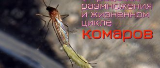
Life cycle
What are the stages of the life cycle of Plasmodium falciparum? Before forming into an adult organism capable of reproduction, such a parasite exists in several intermediate forms. First, human blood becomes infected with microscopic sporangia of an infectious pathogen through a mosquito bite. Then the formation of sexually mature individuals of malarial plasmodium in the body of the intermediate host is observed. The consequence may be the division of mature unicellular organisms or their return to the mosquito, which acts as the main host. In general, the life cycle of Plasmodium involves a periodic change of host.
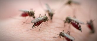
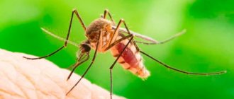
Main owner
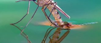
To form a sporogonium capable of reproduction, the malarial plasmodium must enter the body of a female Anopheles mosquito, which lives in the tropical regions of the planet. It is these insects that act as the main host of malarial plasmodium.
Inside the mosquito's body, the microscopic parasite divides, during which independent female and male cells are formed. Each of them has one set of chromosomes. When gametes of individual sexes fuse, cells with a complete chromosome complement are formed. The latter are presented in the form of elongated zygotes. They are extremely mobile, which allows them to penetrate into the tissues of the mosquito’s body, where new incubator cells are formed, covered with a protective membrane. Hundreds of sporangia develop in such containers. After maturation, the walls of the incubator are torn. Single-celled parasites begin to move to the salivary glands of their main host. The malarial plasmodium subsequently seeks to enter the human body.
Intermediate host
The main host of Plasmodium falciparum, which is the mosquito, transmits the causative agents of the disease to humans during a bite. Microscopic parasites quickly spread through the bloodstream throughout the body, concentrating in the liver tissues. Here the stage of asexual reproduction is activated. The result is the formation of so-called merozoites, which infect red blood cells, actively absorb hemoglobin and multiply intensively.
Subsequently, the parasite leaves the red blood cells. It forms digestive vacuoles where nutrients are concentrated. During the processing of food by a pathogenic microorganism, toxins are formed that enter the blood along with the products of its vital activity.
The above stages are repeated periodically. During such cyclical processes, unpleasant symptoms of the disease appear, from which the intermediate host begins to suffer. The malarial plasmodium eventually reaches the apogee of its development, transforming into gametocytes. With the next mosquito bite, the parasites in the presented form penetrate back into the insect’s body, where they return to active sexual reproduction.
Features of the organization of Protozoa
Plasmodium is a representative of the most primitive group of animals - the subkingdom Unicellular or Protozoa. They are characterized by the following signs:
- the body consists of one cell, which performs the functions of the whole organism;
- presence of a core;
- lack of a dense cell wall;
- movement with the help of specialized structures: cilia, pseudopods, flagella;
- the presence of digestive and contractile vacuoles;
- gas exchange through the cell surface;
- sexual and asexual reproduction.
How is malarial plasmodium detected in the blood?
To detect a parasitic infection, a blood sample is examined under a microscope. The sample is taken from a person's finger in a standard way. A smear is then applied to a sterile glass slide. A specialist examines under magnification traces of the presence of malarial plasmodium in any form. The compliance of red blood cell parameters to normal is also monitored. After all, when infected by the malarial plasmodium pathogen, they change not only their shape and size, but also their shade.
Signs of damage to the body by malarial plasmodium
When a tropical mosquito, the final host of Plasmodium falciparum and a vector of infection, infects a person, the disease does not immediately make itself felt. Signs of the parasite's vital activity appear after the incubation period has passed, which often lasts just over a week.
The development of malaria is most acutely felt at the moment when infectious pathogens leave the red blood cells. During this period, the person begins to suffer from fever. A clear sign of the presence of the disease is a significant increase in body temperature, which is accompanied by chills and a feeling of heat. Those infected often fall into delirium caused by severe attacks of headache.
Over time, the above signs of malaria development fade away. Body temperature decreases. Subsequently, the intermediate host of the parasitic infection weakens. However, after some time, the unpleasant symptoms return again. Without proper treatment, destructive changes occur in the structure of the liver and spleen. The body of the infection carrier becomes exhausted. Often, the rapid progression of the disease without adequate help leads to death.
Methods for eliminating infection
The most effective pharmacological agent, the active substances of which slow down the vital processes of malarial plasmodium in the human body, is the drug quinine. The parasitic infection is also low resistant to the substance artemisinin, which is synthesized from the wormwood plant.
To relieve the main symptoms of the disease, the drug primaquine is often prescribed. It is also used for the final destruction of sporangia of the infectious agent in the body.
As you can see, there are several pharmacological drugs to combat Plasmodium falciparum. Despite the availability of drugs on the market, epidemics of the disease are still quite common in tropical regions of the planet. The reason is often the reluctance of the population of underdeveloped countries to use drug treatment, despite the fact that the costs of purchasing effective drugs are often insignificant.
There is also immunity to malaria
- " Innate immunity ". Intermediate carriers with a genetic indicator show some resistance to parasites.
- " Acquired active immunity ". It appears only when a person has had this disease. The level of immunoglobulins increases.
- " Acquired passive immunity" . It appears when ready-made antibodies are introduced into the body.
Sources
- Guide to Medical Entomology / ed. V. P. Derbeneva-Ukhova. – M.: Medicine, 1974. – 359 p.
- Yatusevich, A.I. Little-studied infectious and parasitic diseases of domestic animals: textbook / A.I. Yatusevich, N.N. Androsik. – Mn.: Urajai, 2001. – 331 p.
- Parasitology and invasive diseases of agricultural animals / K.I. Abuladze [and others]. – M.: Agropromizdat, 1990. – 464 p.
- Workshop on parasitology and invasive animal diseases: textbook / A.I. Yatusevich [etc.]. – Mn.: Urajai, 1999. – 279 p.
- Kapustin, V. F. Atlas of blood parasites of animals and ixodid ticks / V. F. Kapustin. – M.: State. ed. agricultural literature, 1955. – 216 p.
- Diagnosis, therapy and prevention of parasitic diseases of horses. UMP for students majoring in veterinary medicine and FPC students. /A.I. Yatusevich, S.I. Stasyukevich, M.P. Sinyakov, V.V. Petrukovich./ Vitebsk, VGAVM, 2011, 60 pp.
- Bulanova-Zakhvatkina, E. M. Armored oribatid mites / E. M. Bulanova-Zakhvatkina. – M.: Higher School, 1967. – 254 p.
- Yatusevich, A.I. Eimeriosis of chickens and its parasitocenological aspects / A.I. Yatusevich, A.V. Sandul, V.N. Gisco. – Vitebsk: VGAVM, 2009. – 249 p.
- Parasitology and invasive diseases of farm animals / K. I. Abuladze, N. V. Demidov, A. A. Napoklonov, etc.; edited by K. I. Abuladze. – 3rd ed. – M.: Agropromizdat, 1990. – 464 p.
- Balashov, Yu. S. Blood-sucking ticks (Ixodoidea) - carriers of human and animal diseases / Yu. S. Balashov. – L.: Nauka, 1967. – 320 p.
- Determinant of helminths of hunting mammals in Belarus: monograph / A.I. Yatusevich [and others]. – Vitebsk: VGAVM, 2010. – 228 p.
- Veterinary and sanitary measures for the prevention and elimination of parasitic diseases of animals: guidelines / I.N. Dubina [and others]. – Vitebsk: VGAVM, 2008. – 51 p.
- Dubina, I.N. Veterinary and sanitary rules for the prevention and elimination of animal cestodiasis: approved. GUV MASHIP RB / I.N. Dubina, A.I. Yatusevich. – Vitebsk: VGAVM, 2007. – 10 p.
- Yatusevich, A.I., Parasitic diseases of birds / A.I. Yatusevich, B.Ya. Birman, T.G. Nikulin. – Mn.: Publishing house Polibig, 2001.
- Olenev, N. O. Scabies mites / N. O. Olenev. – L.: Ed. Academician Sciences of the USSR, 1932. – 64 p.
- Kuznetsov, M.I. Anoplocephalatosis of ruminants / M.I. Kuznetsov. – M.: Kolos, 1972.
- Stepanova, N.I. Protozoal diseases of animals / N.I. Stepanova. – M.-L.: Kolos, 1982.
- Adaptation processes and parasitosis of animals: monograph / A.I. Yatusevich [and others]. – Vitebsk: OU VGAVM, 2006. – 404 p.
- Yatusevich A.I., Poorly studied infectious and invasive diseases of domestic animals / A.I. Yatusevich, N.N. Androsik. – Mn.: Urajai, 2001. – 332 p.
- Dubinina, M. N. Parasitological studies of birds / M. N. Dubinina. – L.: Nauka, 1971. – 139 p.
- Savchenko, S.V. Prevention and treatment of balantidioesophagostomotic infestation in pigs / S.V. Savchenko, V.F. Savchenko. – Vitebsk: VGAVM, 2010. – 194 p.
- Theoretical and practical principles of the use of medicinal plants for parasitic diseases of animals / A.I. Yatusevich [and others]. – Vitebsk: VGAVM, 2008. – 75 p.
- Dubinin, V. B. Scabies mites. Biology, harm in agriculture, measures to prevent and combat them / V. B. Dubinin. -M.: Soviet Science, 1954. – 173 p.
- Yatusevich, A.I. Arachnoenthomoses of domestic ruminants and single-hoofed animals: monograph / A.I. Yatusevich [and others]. – Vitebsk: VGAVM, 2006. – 213 p.
- Subbotin, A.M. Sparganosis of wild boar: differential diagnosis and improvement of measures to combat it in the conditions of the Republic of Belarus: method. instructions / A.M. Subbotin, N.F. Karasev, V.F. Litvinov. – Vitebsk: VGAVM, 2009. – 24 p.
- Yatusevich, A.I. Eimeriosis nutria: monograph / A.I. Yatusevich, V.A. Forget it. – Vitebsk: OU VGAVM, 2006. – 87 p.
- Parasitology and invasive animal diseases / M.Sh. Akbaev [and others]. – M.: Kolos, 2000. – 743 p.
- Yatusevich, A.I. The pratazoin sicknesses of the rural and gaspdarchy lived. – Mn.: Urajai, 1993. – 174 p.
- Animal cryptosporidiosis (Recommendations for diagnosis, therapy and prevention). Approved by the Main Directorate of the Ministry of Agriculture and Public Administration of the Republic of Belarus on July 25, 2000. Authors: Yatusevich A.I., Savchenko V.F., Nesterovich S.G. and etc.
- Balashov, Yu. S. Blood-sucking arthropods and rickettsia / Yu. S. Balashov, A. B. Deiter. – L.: Nauka, 1973. – 251 p.
- Parasitology and invasive animal diseases / M. Sh. Ak-baev, A. A. Vodyanov, N. E. Kosminkov, etc.; edited by M. Sh. Akbaeva. – M.: Kolos, 2002. – 743 p.
- Recommendations for the fight against helminthiasis in horses / A.I. Yatusevich [and others]. – Vitebsk: VGAVM, 2008. – 15 p.
- Dubinin, V. B. Scabies mites. Biology, harm in agriculture, measures to prevent and combat them / V. B. Dubinin. -M.: Soviet Science, 1954. – 173 p.
- Agrinsky, N. I. Insects and mites that harm farm animals / N. I. Agrinsky. – M.: Selkhozizdat, 1962. – 288 p.
- Sokolova, T. V. Scabies / T. V. Sokolova, R. F. Fedorovskaya, A. B. Lange. – M.: Medicine, 1989. – 176 p.
- Yatusevich, A.I. Veterinary and medical parasitology: (Encyclopedic reference book) / A.I. Yatusevich, I.V. Rachkovskaya, V.M. Kaplich. – M.: Medical literature, 2001. – 309 p.
- Yatusevich, A.I. Eimeriosis of chickens: monograph / A.I. Yatusevich, V.N. Gisco. – Vitebsk: OU VGAVM, 2007. – 103 p.
- Karasev, N.F. Larval cestodiasis of animals / N.F. Karasev, T.G. Nikulin, N.K. Slepnev. – Mn.: Urajai, 1989. – 111 p.
- Kapustin, V.F. Atlas of helminths of farm animals. – M.: Selkhozgiz, 1953. – 139 p.
- Dubina, I.N. Methodological guidelines for diagnosing helminthiasis of carnivores: approved. GUV MASHIP RB / I.N. Club. – Vitebsk: VGAVM, 2008. – 32 p.
- Yatusevich, A.I. Helminth infections of cattle and measures to combat them under environmental pressure / A.I. Yatusevich, R.N. Protasovitskaya. – Vitebsk: VGAVM, 2010. – 160 p.
- Veterinary laboratory practice. T. 2. – M.: Selkhozizdat, 1963. – 432 p.
- Orlov, I.V. Workshop on veterinary parasitology / I.V. Orlov, N.I. Agrinsky, S.N. Nikolsky. – M.: Selkhozizdat, 1962. – 319 p.
- Practical parasitology / ed. D. V. Vinogradov-Volzhinsky. – L.: Medicine, 1977. – 304 p.
- Antiparasitic drugs for dogs and cats: educational method. allowance / A.I. Yatusevich [and others]. – Vitebsk: VGAVM, 2009. – 48 p.
- Shershen, G.G. Methodological recommendations for identifying natural foci of piroplasmosis in animals in Belarus / G.G. Shershen, N.F. Karasev. – Vitebsk: VGAVM, 2009. – 32 p.
- Midges and bovine simulidotoxicosis. / Skulovets M.V /-2007
- Parasitic diseases of horses / A.I. Yatusevich [and others]. – Mn., 1999. – 78 p.
- Kaplich, V.M. Gnus and measures to combat it in Belarus / V.M. Kaplich, A.I. Yatusevich, M.V. Skulovets. – Mn.: Urajai, 1994. – 120 p.
- Dubina, I.N. Guidelines for the diagnosis of larval cestodiasis in animals / I.N. Club. – Vitebsk: VGAVM, 2008. – 39 p.
- Diseases of agricultural animals / P.A. Krasochko [and others]; under. ed. P.A. Beautiful. – Mn.: Businessofset, 2005. – 800 p.
Prevention of Plasmodium falciparum infection
The main preventive measure to prevent the spread of malaria is the destruction of the tropical Anopheles mosquito, which acts as the final host and carrier of the infection. Without such insects, the microscopic parasite simply does not have the opportunity to begin and complete its life cycle.
In order to protect against the bites of malaria mosquitoes, they often resort to the use of concentrated repellents. When staying in potentially dangerous areas, it is recommended to wear clothes that cover exposed areas of the body and use mosquito nets.
If you plan to travel to places where there is a possibility of infection with malarial plasmodium, prophylactic use of medications such as Chloroquine, Delagil, Rezoquin, which are available in tablets, is required. The action of these drugs is based on slowing down the synthesis of nucleic acids in the body, which leads to inhibition of the vital activity of the infectious agent. It is advisable to take pills for preventive purposes for another month after leaving a potentially dangerous region.