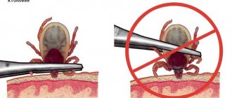
What is Larva Migratory Syndrome?
The content of the article
Based on the developmental characteristics of the parasite, two types of hosts are distinguished: intermediate and final.
- The host containing a sexually mature worm is called final.
- The host containing the developing worm larva is intermediate.
Helminths, entering the host’s body, use its nutrients, are toxic to the nervous system, poison internal organs and therefore can cause various changes in the body.
Animal nematode larvae that enter the human body can cause larva migrans syndrome. Since humans are an intermediate host, pathogen larvae do not grow or mature in the human body, but can migrate through tissues and cause inflammation.
This syndrome can be caused by several types of helminths. However, it is most often caused by:
- round dog worms Toxocara canis;
- less commonly, cat roundworms Toxocara cati;
- or Toxocara mystax larvae.
The disease caused by Toxocara larvae entering a living organism is called toxocariasis. This is a dangerous chronic disease, it lasts a year or more with exacerbations. The prevalence of the infection is associated with the infection of dogs and cats and the contamination of the environment with their feces.
Types of helminths
There are many types of internal parasites in the world, but the most common types of worms in dogs are the following:
- Nematodes (roundworms).
- Trematodes (flukes).
- Cestodes (tape).
According to the place of localization (parasitism), parasites are: liver, pulmonary, intestinal, subcutaneous, cardiac. The most common and frequently diagnosed are roundworms in dogs, Toxascaris leonina or canine roundworm, a nematode that reaches a length of up to 20 cm and affects cats and humans.
Toxocariasis - migration syndrome
The larvae penetrate the skin. Toxocara eggs are found in large quantities in the soil. Toxocara larvae are most often found in children, affecting 2% to 10% of those infected. The incidence is the same in boys and girls. The age of sick children is from 1 to 7 years.
Infection of children with the disease is caused by:
- failure to comply with hygiene requirements;
- playing in the sandbox or with animals.
It has been established that in children aged 1–6 years, geophagia manifests itself as a behavioral disorder in 2–10%. This disorder is important because at the onset of intraocular larval migration syndrome, 40% of patients have this behavioral pathology.
The disease is less common among adults. In Western Europe, 2–5% are affected. Healthy adults have antibodies to Toxocara.
The incidence of toxocariasis also depends on the place of residence. Toxic pathogens are more common in tropical and temperate climates, and in warm countries their number reaches 86-93%. Infectiousness is much higher among rural populations (14-37%) compared to urban populations.
According to various countries, infection of Toxocara eggs in courtyards, sandboxes, beaches and other public places ranges from 10% to 30%. This problem is associated with an increase in the number of dogs and cats left unattended and non-compliance with the deworming schedule for pets.
Pathogenesis
Pathogenesis
The final host of roundworms is a member of the dog and cat family, only in which the worm can grow, mature and lay eggs. When a worm enters a dog’s body, it parasitizes the intestines and feeds on the intestinal contents.
Dogs become infected with Toxocara by eating worm eggs in the soil. In the intestine, the larva released from the egg migrates with the blood to the lungs, bites the alveoli and enters the nasopharynx and mouth through the bronchi. This part of the migration, the airways, is called the tracheal stage of migration.
Later, the larvae enter the dog's intestines again through the esophagus, where they develop into adult worms that lay eggs. An adult female Toxocara produces about 200-250 thousand individuals. The remaining larvae penetrate the tissues and are distributed throughout the tissues of the body.
Studies have shown that in the female body, some of the larvae can remain in the tissues for a long time. In the last weeks of a dog's pregnancy, the larvae can become active and infect future puppies due to various hormones. Puppies can also become infected while feeding. Such an infected bitch can give birth to up to four puppies in a litter infected with Toxocara canis, without re-infection.
Thus, the main source of toxocara in humans is puppies under 3 months of age. Therefore, deworming pregnant bitches and all pets as needed is an important part of prevention.
With the dog's feces, the eggs fall into the soil and begin to mature, becoming an infection. Soil and related environmental conditions are important for egg development:
- air temperature (16-30 o C);
- air and soil humidity;
- light.
In soil, eggs mature in 2–3 weeks. Under the wrong conditions, the eggs go into a state of suspended animation and thus can survive the winter. Eggs can remain viable in soil for 8 years or more.
Humans are occasional intermediate hosts for this parasite and become infected with toxocariasis through oral feces. Unlike animals, larvae that do not enter the human body do not reproduce or grow, so people cannot become infected with them. Most often, the larva is transmitted through dirty hands when eating unwashed berries, fruits, and vegetables. It is also possible to become infected by eating contaminated and undercooked chicken, beef or lamb. Children can become infected while playing in the sandbox.
People cannot become infected with toxocariasis from a dog through contact, since the eggs must enter the soil and mature. However, care must be taken as dogs may carry mature eggs on their fur or tongue.
Mature Toxocara eggs enter the gastrointestinal tract through the mouth. In the human stomach, the eggshell dissolves, the larva is released and migrates towards the small intestine. It penetrates the intestinal mucosa and enters the blood and lymphatic vessels. Together with blood and lymph, the larvae enter the organs:
- liver;
- heart;
- lungs;
- kidneys;
- pancreas;
- eyes, etc.
It has been established that Toxocara cati larvae mainly migrate to the muscles, liver and lungs, while Toxocara canis larvae are often found in the central nervous system.
The larvae are encapsulated in tissues for a long period of time, after which the capsules disintegrate and the larvae are released. At this time, they become active and begin to migrate, irritating and damaging tissue. The movement of the larvae causes an immune tissue response and an allergic reaction.
This movement of larvae is called larval migratory syndrome. The condition can only be caused by the larval stage of the parasite and not by the adult worm or eggs. Depending on the location of the larvae, the syndrome is divided into two types:
- visceral;
- intraocular.
Symptoms of infection
This disease does not have pronounced symptoms, which can clearly indicate the presence of parasites of this particular species. In most cats and dogs, the disease manifests itself as vague symptoms, the intensity of which depends on the type of lesion and the time the parasites live in the body.
In the cutaneous form, everything is usually limited to the following symptoms:
- in the place where the worms have settled, severe itching develops, a rash and dermatitis appear. The animal itches vigorously, which can lead to thinning and hair loss, as well as swelling;
- If an infection gets into the wounds when scratching, inflammation may develop, which can mask the presence of helminthic infestation.
Signs of pulmonary-cardiac form:
- Heavy breathing, shortness of breath.
- The appearance of sputum with blood.
- Cough, not expressed, not strong.
- Swelling on the paws.
- Lethargy, weakness.
- Poor appetite.
- Apathy.
- Fast fatiguability.
- Fever, low-grade temperature.
- Some animals have glomerulonephritis, renal and liver failure. Against this background, ascites may appear in a dog; the symptoms are described here.
Helminths can penetrate not only the heart and lungs. Their presence can be detected in the brain and spinal cord, in the eyes and in the abdominal cavity.
Characteristic signs are weight loss, ascites, especially with cardiac problems, cyanosis of the mucous membranes, severe weakness and fainting, moist rales and many other manifestations.
Typically, pronounced signs appear in the later stages of the disease, when the larvae reach the adult stage and die. The decay products of parasites, especially when there are large numbers of them, can cause severe intoxication in the dog’s body.
Symptoms and treatment of the disease are interrelated, but an accurate diagnosis must first be established in order to select the correct medications for a particular animal.
Clinic
Clinical symptoms of toxocariasis vary widely and depend on the organs and tissues affected by the migratory larvae. There may also be no symptoms.
The course of the disease depends on:
- number of larvae;
- affected organ;
- general resistance of the body;
- re-infection rates.
Since migratory larvae cause allergic inflammation, the severity of the disease also depends on the body's allergic reaction to the toxocarial larvae. In patients with atopic allergic disease, this infection is usually more severe.
Specific antibodies cause an inflammatory response to the toxocarsecretory (TES) antigen and the allergic component TBA-1. These antigens activate CD4+ cells and they differentiate into Th1 and Th2 cells. Interleukin-2 (IL-2) and γ-interferon (γIFN) secreted by Th1, interleukin-4 (IL-4) and interleukin-5 (IL-5) induce additional immune responses.
IL-4 promotes the proliferation and differentiation of B cells into plasma cells; when exposed to IL-2, IL-4, IL-5 and γIFN, they begin to synthesize antibodies of various isotypes. IL-5, in turn, promotes eosinophil differentiation and adhesion.
Epithelial inflammatory cells surround the larvae and form specific eosinophilic granulomas. It is the localization of granulomas that determines the clinical course of the disease.
There are different forms of visceral and ocular (intraocular) larva migrans syndrome. This classification is greatly simplified since one of these forms is always possible. And detection of toxocariasis of the eye and examination of internal organs usually reveal varying degrees of damage to internal organs.
Visceral migratory larval syndrome
Toxicosis can affect various organs:
- skin;
- lungs;
- liver;
- joints;
- heart;
- eyes;
- brain.
Thus, the syndrome manifests itself with various symptoms. However, the most common areas affected are the respiratory tract, making signs of upper respiratory tract inflammation one of the most common.
Unexpected manifestations of visceral larval migration syndrome are also possible after the sudden migration of a large number of larvae:
- acute pneumonia;
- eosinophilic meningoencephalitis.
This syndrome may increase the risk of death if the infection is accompanied by an abnormal immune response or when there is extensive migration of larvae into the myocardium or central nervous system.
Development cycle
The female flies up to the future “incubator” and literally injects the larvae into the affected area of the skin.
Each “injection” contains at least 10-15 individuals. The larvae quickly burrow into the wound, actively feed and molt twice. After about five days (depending on the ambient temperature), they crawl out of their cozy nest, fall onto the soil, burrow into it, and pupate. After one to three weeks, the imago (that is, the adult stage) emerges.
Interesting!
Adult flies are most active at an ambient temperature of 20-27 degrees Celsius. If it gets hotter, they find shade and fall into a kind of torpor.
Intraocular larva migrans syndrome
This type of syndrome occurs when toxocariasis larvae enter the tissues of the eye and orbit through the central retinal artery and dentate arteries. Ocular toxocariasis is classified according to the 1971 Wilkinson and Welch classification according to the developing clinic. Only now the classification has become more precise as more is known about the syndromes.
Classification:
- Chronic endophthalmitis (vitreous rays, membranes, granulomas; posterior uveitis is pronounced; visual acuity is sharply reduced);
- Posterior granulomatous chorioretinitis (in the posterior pole of the eye, in the macula, on the optic nerve papilla or adjacent to these retinal structures there are 1-2 white lesions; usually visual acuity is reduced);
- Peripheral granulomatous chorioretinitis (white formations covered with vitreous connective tissue, penetrating the vitreous located on the periphery of the retina; visual acuity usually does not change).
In rare cases, ocular toxocariasis may manifest as:
- neuritis;
- neuroretinitis;
- localized vitreous abscess;
- iridocyclitis;
- larval keratoconjunctivitis.
If toxocariatic larvae enter the tissue in front of the orbital septum, this causes inflammation of the anterior septum cell: the eyelids and conjunctiva become swollen and red. When the larvae enter the orbital septum, the orbit becomes inflamed.
Cellular inflammation:
- swelling and redness of the joints and eyelids;
- painful eye movements;
- doubling;
- ophthalmoplegia;
- inversion;
- Optic neuropathy.
Most often, one eye is affected, and visual changes directly depend on the location of the lesion. Ocular toxocariasis is often complicated by:
- retinal circulatory disorders;
- retinal detachment;
- abscess of the eye;
- secondary glaucoma or cataract.
Diagnostics
Diagnosis
Diagnosis of all helminth infections is based on taking an accurate history. Epidemiological aspects are very important, since the risk of developing a particular infectious disease is closely related to:
- profession;
- rest;
- travel to certain endemic areas.
But they alone are not enough.
Of course, diagnosing toxocariasis is not easy, because people do not excrete toxocariasis worms or eggs, and it is also difficult to detect migrating larvae in biopsy material. Therefore, to confirm the diagnosis, instrumental and laboratory tests are always carried out:
- Complete blood count
: leukocytosis, persistent eosinophilia, anemia, thrombocytosis. Eosinophilia is reported to be more common in children than in adults; - Immunological tests.
More common: hypergammaglobulinemia. The level of IgE gamaglobulin increases. Increased isohemagglutinin titers for blood group A and B antigens. Antibodies to Toxocara are found in the vitreous body (90% of cases); - Linked immunosorbent assay
. Toxic secretory (TES) antigens are detected using direct ELISA. For toxocrosis, the specificity of the ELISA assay is 92–95% and the sensitivity is 73–75% when using a titer of 1:32 for diagnosis (4, 14). Indirect ELISA is used to detect antibodies (IgE, IgG and IgM) to TES toxocarial antigens. This method detects antibodies from 78 to 92%. cases; - Instrumental research methods
. In the visceral form of the syndrome, ultrasound or computed tomography reveals an enlarged liver, and intraocular examination reveals granulomas or abscesses in the tissues of the eye. Granulomas in the cortex or subcortex can also be seen on CT scans. - BMR Study.
Shows changes in the brain. Protein exudate in T1 and T2 modes may be more pronounced, and the potential difference may be visible. Gadolinium testing improves detection of abscess. Toxicara larvae can be identified by organ biopsy. It has been noted that larvae are extremely rare in biopsy material, but if they are not found, the diagnosis cannot be excluded; - Histological examination
. Multiple eosinophilic abscesses, allergic-type granulomas in damaged organs, and tissue necrosis around macrophages and lymphocytes were detected.
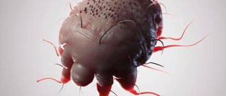
What do parasites look like?
The presence of lice eaters on a dog’s body provokes a dangerous disease called trichodectosis. And before you start fighting parasites, you need to know what they look like.
The lice eater is a small insect that parasitizes the body of warm-blooded animals. Its flat, flattened body does not exceed 2 mm in length and most often has a grayish-transparent or pale yellow tint. Therefore, detecting a pest is not always easy. Externally, lice eaters are very similar to lice. Their distinguishing feature is their head. In wingless insects it is quadrangular in shape and much larger than the chest. Lice eaters reproduce quite actively. A female lice eater can lay 60-70 eggs, which are firmly attached to the dog’s hair using an adhesive mass, making these eggs difficult to wash off with water and comb out. After 5-10 days, larvae emerge from the laid eggs, which molt at least three times before becoming sexually mature. The entire development cycle lasts about a month.
Below you can see what lice eaters look like in dogs in the photo.
Pathogenesis of treatment
Detoxification therapy is used, including intravenous infusions of electrolytes. To restore dehydration, a 20% mannitol solution is administered intravenously. Intravenous or intramuscular administration of furosemide until the desired result is achieved. Nonsteroidal anti-inflammatory drugs (NSAIDs) are used to reduce the inflammatory response by reducing the activity of cyclooxygenase and the synthesis of prostaglandins and leukotrienes.
Damage to the central nervous system in larvae can lead to agitation and is suppressed by sedatives. For the form of ocular toxocariasis, all of the above drugs are prescribed. In severe cases or in the presence of complications, surgical treatment may be used:
- vitrectomy;
- cataract surgery;
- laser photocoagulation;
- enucleation of the eye.
What danger do lice eaters pose to dogs?
Like other ectopaparis (fleas, ticks, lice), lice eaters are carriers of tapeworms and a variety of infectious diseases. When lice eaters settle on a dog, they cause severe itching (itching in dogs), as a result of which numerous scratches, irritations, and dermatitis appear on the skin (dermatitis in dogs). The presence of an ectoparasite leads to a decrease in immunity, and anemia may develop.
Prevention
Toxicar eggs pollute the environment, so it is very important to follow the basic rules:
- personal hygiene;
- deworming of pets;
- clean environment;
- security of the children's playground.
Personal hygiene: every time you return from the field, you must wash your hands after stroking sand, soil and petting pets. Also, do not allow dogs to lick their faces or utensils used by people. Eat only washed berries, fruits, and vegetables.
Pets need to be dewormed promptly. Because puppies and kittens can become infected by transplantation or through breast milk. Deworming should begin after 3 weeks and continue every 2 to 4 weeks. Up to 6 months Puppies over 6 months of age are recommended to be dewormed every month. For adult dogs every 3 months. Bitches should be additionally dewormed 2 weeks before mating.
When going for a walk with your pet, be sure to pack bags to collect feces. This will prevent animal feces from polluting the environment. Pets should be avoided on playgrounds. When children are not playing, sandboxes should be covered. The sand in the boxes also needs to be changed regularly. The main and biggest problem is stray dogs and cats, so catching them and transporting them to shelters is an integral part of professional tactics.
Is it worth calling a doctor to your home to get help on how to remove maggots from a dog?
How to prepare for the doctor's arrival
Having a doctor visit your home can save you a lot of time.
, which can be spent usefully, for example, inspecting the rear hole of the animal for cleanliness. You should also carefully prepare for the doctor’s visit. Prepare a clean towel, wet wipes, and a large, wide surface covered with a sterile sheet. When the doctor arrives, you should remain calm and not panic. Dogs are excellent psychologists and they detect the slightest change in their owner’s mood. Panic is also easily transmitted to them, and an agitated animal is quite difficult to examine. Which doctor should I call? First of all, you should invite a veterinarian who will conduct a general examination of the animal. By agreement with the owner, a dermatologist from our veterinary center will be sent.
- You can further familiarize yourself with the list of our doctors who are ready to help on the first call:
- classical doctor;
- dermatologist;
- surgeon;
- dentist;
- nephrologist;
- neurologist;
- ophthalmologist;
- gynecologist for bitches;
- ratologist (specialist in rodents and guinea pigs).
If you use such a convenient service as a veterinarian at home at least once, you will no longer want to come to the veterinary center on your own.