The manifestation of unpleasant symptoms on the skin is very often the result of damage by skin mites. This type of disease greatly worries many people who have encountered this type of tick. Skin mites on the face cause especially a lot of discomfort.
Infection occurs very quickly and spreads throughout the entire face, including the hair growth area. A large number of external environmental factors and problems with the functioning of internal organs can lead to the appearance of this type of skin disease.
Skin mites do not disappear on their own; eliminating them requires the use of special medications and timely examination. If left untreated, the condition may deteriorate and a large area of the body may be affected.
What is demodex?
The appearance of skin mites is referred to in medicine as demodex. It is impossible to see such an organism with the naked eye; the skin mite is microscopic in size and is most often localized in the face, neck, chest and shoulders.
Features of Demodex:
- The largest number of skin mites is found in the sebaceous glands;
- In humans, skin mites can live for a long time in the upper layers of the skin and not cause any unpleasant symptoms;
- After weakening of the immune system or in the event of a violation of the protective functions of the skin, the tick has the ability to penetrate into the deep layers and form purulent, inflamed formations;
- Women most often suffer from this type of skin disease;
- To identify the type of disease, it is necessary to undergo a special type of diagnosis, during which the source of unpleasant symptoms will be identified.
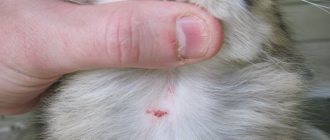
Externally on the skin, a skin mite most often appears in the form of pimples with the presence of pus or subcutaneous nodules, which after some time become inflamed and increase in size.
What does a tick look like?
Examining the components of inflamed formations under a microscope, you can see:
- The demodex mite is a type of glandular acne, the reproduction of which occurs in the sebaceous glands;
- Outwardly it resembles a thin transparent worm;
- The tick's shell has no color, but is dense;
- The size of the tick is on average 0.4 mm;
- The skin mite moves with the help of scales;
- Demodex feeds on skin cells and subcutaneous fat.
Demodex most often exhibits its activity at night or during the development of diseases that reduce immunity.
Life cycle
The entire life course of a worm-like mite can be divided into two periods of existence:
- intradermal (maturation and reproduction)
- extracutaneous or cutaneous
The mite, fixed on the walls of the excretory ducts, reaches a sexually mature state, is fertilized, and lays eggs. This concludes the ripening period.
Then a cutaneous, metamorphic period takes place outside. The eggs reach the surface of the dermis with a current of sebum. The larvae that emerge from them settle under the scales of the epidermis, in the mouths of the hair follicles. Molting (metamorphosis) takes place there - protonymph and teleonymph.
Subsequently, adults roll down the excretory duct of the sebaceous glands, and the cycle begins again. Fertilization is internal; individuals mate at the mouth of the follicle. In total, the life cycle takes 15-25 days.
Source and causes of occurrence
The mite can live on the skin and hair of almost every second person.
However, under normal conditions of functioning of internal organs, further spread of the tick does not occur. This disease is also called demodicosis.
The causes of the disease are as follows:
- Disruption of the digestive organs;
- Frequent nervous breakdowns;
- Insufficient or excessive skin care;
- Incorrectly selected cosmetics;
- Excessive use of saunas and solariums;
- Excessive coffee consumption and bad habits;
- Spicy food;
- Decreased functioning of the endocrine gland.
You can also become infected with a tick through close contact with a sick person, for example, sharing a bed or sharing cosmetics. The disease cannot be transmitted by air or other types of contact.
Circumstances that determine the effectiveness of treatment
As mentioned earlier, getting rid of skin mites is not so easy, so the effectiveness of treatment depends on many factors, such as endurance, patience, following all the instructions of the attending physician, etc. In addition, you need to adhere to a number of rules, such as:
- A healthy diet without fatty, spicy, salty and fried foods, with limited carbohydrate intake. The diet should include fermented milk products, fresh vegetables and fruits. You will have to stop smoking and drinking alcohol.
- You will also have to refuse to visit the bathhouse, sauna and solarium.
- Personalize all accessories such as cosmetics, clothes and shoes.
- Avoid using down pillows and blankets, as ticks can easily live in them, and they can cause a relapse.
- Be sure to iron bedding after washing.
- You should not squeeze out pimples that appear as a result of mites on your own.
- You should only use cold water to wash your face.
- It is forbidden to self-medicate, as this disease can become chronic.
Demodicosis is a very serious disease. The sooner you start treatment, the sooner you will be able to get rid of subcutaneous mites. When the disease is advanced, treatment can take six months, or even more.
Therefore, it is better not to start treatment with “grandmother’s” remedies, which can relieve the feeling of discomfort, but they will not kill the ticks, and they will continue their vital functions, although not as actively.
Only chemicals and proper treatment, with the correct alternation of different drugs, will help you get rid of ticks. In other words, at the first suspicion of a subcutaneous mite, it is better to immediately go to a dermatologist.
Prognosis and complications
Skin with demodicosis has a condition that contributes to the development of other dermatological diseases. In this case, the symptoms manifest themselves much more strongly. With demodicosis of eyelashes, the development of conjunctivitis and blepharitis is possible.
Scratching and regular mechanical damage associated with the desire of patients to get rid of rashes can provoke a pustular infection that affects many parts of the body.
Complications include nervousness and isolation. Acne that appears all over the body significantly spoils the appearance and many are unable to lead a normal life. In some cases, this leads to depression, nervous breakdowns and psychosis. People diagnosed with demodicosis often have endocrinological disorders, chronic gastrointestinal diseases, or foci of chronic infection.
Preventive measures
In order not to provoke the disease again after recovery, you must adhere to simple rules of hygiene and be careful about your diet:
By adhering to these simple rules, you can not be afraid of the resumption of active actions of subcutaneous mites.
Source
Signs and symptoms of the disease
Very often, mite infestation is confused with acne and the correct treatment is not used.
Demodex manifests itself with the following first signs:
- The skin becomes oilier and visual enlargement of pores is observed;
- Formation of places on the skin with an increased level of shine;
- Redness and swelling of the skin;
- Itching at night;
- The appearance of scales on the eyelashes, which can lead to burning and itching.
After infection, the following unpleasant symptoms occur:
- Inflammation and swelling of certain areas of the skin;
- The appearance of red bumps;
- Hair loss and itching on the scalp;
- The appearance of pimples in large numbers with purulent inclusions;
- Puffiness of the eyes;
- Formation of ulcers;
- The appearance of plaque in the area of hair growth.
Symptoms may appear in isolated cases or with a high level of distribution in healthy areas of the skin.
Folk remedies
Remember that folk recipes can help with drug treatment. Separately, they are ineffective and one should not refuse the help of doctors, as this will aggravate the situation.
Celandine juice is quite effective, but it is not suitable for everyone, since it can cause allergic reactions. It is used in the treatment of scabies. To use it, you need to dilute the juice with water in a ratio of 1:2, then wipe the rash with a cotton swab.
For demodicosis, tea tree oil is used, which helps relieve itching, relieve burning and improve skin condition. To do this, you need to take body cream and add a few drops of oil to it. Juniper infusion is also used.
To prepare it you will need dry berries that need to be chopped. One tablespoon of crushed berries is poured into a glass of boiling water and left for 5 hours, then filtered and used for rubbing. To increase effectiveness, before wiping, you need to wipe the skin with calendula infusion, and then use the infusion as a compress, which should be kept for no more than 20 minutes.
In addition to drug treatment and the use of traditional methods, a very important point in therapy is proper nutrition.
Stages of development
Depending on the number of mites and the degree of their progression in the deep layers of the epidermis, different stages of the disease are distinguished.
Such as:
- The prodromal stage is the first stage when a tick infection has just occurred. During this period, practically no unpleasant symptoms are felt; the disease can only be identified using special diagnostic methods. This period is considered the most optimal for the complete elimination of the skin disease. The very first symptoms are slight redness of the skin in the areas where the sebaceous glands are located;
- Erymatous stage - the disease begins to progress and a person can already visually observe the first visible symptoms, such as swelling and the formation of purulent pimples. This stage can be treated without the use of complex therapy;
- The papulopustular stage is a more complex stage of the development of the disease, which manifests itself in the form of a large number of pimples, accompanied by specific itching and inflammation of the skin. Treatment requires special examination and observation by the attending physician;
- The hypertrophic stage is considered the most complex course of the disease, which is manifested by large inflamed formations containing pus. At this stage, it is almost impossible to eliminate the disease.
To prevent the development of complex stages of the disease, it is recommended to seek help in a timely manner and not self-medicate.
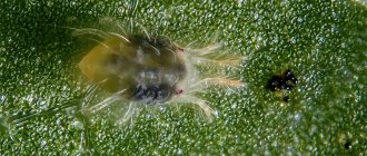
What are ticks?
Ticks are arthropod invertebrate animals from the class of arachnids. Now there are about 50 thousand species.
Thanks to their microscopic size, they were able to easily adapt to their environment.
Some of them parasitize humans and animals. Others, for example, oribatid mites, being saprophages, live in the ground and feed on the products of decomposition of vegetation, mold, and carrion. They are beneficial because they participate in the formation of humus. They do not pose a danger to humans, but if they enter their home, they can cause allergies.
Ticks cause a number of diseases in humans called acariases. There are many of them. These include: tick-borne encephalitis, scabies, demodicosis, allergic manifestations, various dermatitis.
In addition, arthropods are carriers of many infectious pathologies, including, for example, Lyme disease, piroplasmosis, bartonellosis, and tularemia.
According to the method of parasitism, ticks are divided into 2 categories: cutaneous and intradermal. The latter include microscopic parasites whose entire life cycle takes place in the human body. These are the following types of ticks:
- sarcoptoid;
- demodexes.
External species of creatures, or ectoparasites, are found on the skin of humans or animals.
Ticks on the human body feed on blood, plasma, and epithelial cells. Arthropod parasites land on the skin to obtain fresh blood. Then they return to their usual environment, where reproduction occurs. They can be seen with the naked eye: they are up to 5 mm long, and some even up to 1 cm. Skin mites that can parasitize humans include cheyletiella and the ixodid tick.
Ticks feed on blood, lymph and skin
The usual route of infection with ticks is contact with an infected person or animal, the use of shared hygiene items, clothing that belongs to the patient, and walks in nature.
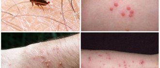
Common symptoms of ticks in humans are: itching, often worsening at night, redness of the skin, and rash on the body.
What triggers the growth of subcutaneous mites?
The following factors lead to the rapid growth and reproduction of ticks:
- Improper skin care;
- Insufficient intake of vitamins;
- Disease of internal organs;
- Lack of treatment for skin mites;
- Use of antibiotics;
- Dysbacteriosis;
- External negative factors.
In addition, increased activity of the sebaceous glands increases the growth of the mite, which can occur as a result of excessive use of products containing alcohol.
Routes of infection
Subcutaneous mites are not always activated in the human body due to health problems or poor immunity; in some cases, accidental infection with these parasites is possible. Such infection can occur in the following ways:
- Personal hygiene products, cosmetics or bedding if they were used by an infected person.
- Contact with the sebaceous glands or hair of an infected person.
- Using non-sterile instruments in hairdressing or massage parlors.
Diagnostics
To identify the stage of development, it is necessary to carry out special diagnostic methods:
- Scraping from the site of inflammation - using a scalpel, carefully take a scraping from the affected areas of the skin;
- Eyelash test - several eyelashes from both eyes are taken for analysis;
- Using adhesive tape , which is glued to the affected area for a certain time, after which it is checked for the presence of ticks.
After receiving the tests, the doctor prescribes the necessary treatment and diet to improve the general condition of the body.
O.V. Kalinina, K.N. Pustovaya, V.I. Nozdrin
JSC "Retinoids", Moscow
Summary
The literature review examines the main aspects of the available information about Demodex mites in humans. The article presents data on the classification, prevalence, structure, types, life cycle, methods of their detection and diseases associated with them.
Keywords:
Demodex mites, rosacea, morbidity, diagnosis.
General information about Demodex mites
Demodex mites are small parasites belonging, according to taxonomic classification, to the phylum Arthropoda, class Arachnida, order Acariformes, family Demodicidae, genus Demodex. More than 140 species of ticks have been described, found both in humans and in various mammals (rodents, domestic animals, small and cattle). Currently, the morphology of most Demodex mites has been well studied, and the generalized damage they cause for animals is a potentially dangerous condition, if untreated, fatal, which is associated with intensive proliferation of mites and the addition of a secondary infection [2, 3]. Despite the fact that ticks are highly species-specific, there are reports in the literature of cross-infection with these ticks between humans and animals [4, 5]. Some authors are studying the issue of the relationship between mites and the host organism, although the issue of Demodex residence continues to remain unclear [6, 7]. In the specialized literature, there is a description of an increasing number of epidemiological and clinical studies that indicate the important role of Demodex mites in rosacea and pityriasis [8, 9], as well as seborrheic and perioral dermatitis, blepharitis, alopecia and other lesions of the skin and its appendages [10, 11, 12]. There are observations of an increase in the number of mites in rosacea under long-term use of glucocorticosteroids [15, 16]. It is assumed that the development of neoplasms of the skin and its appendages promotes the production of protease and cytokines, which leads to a decrease in immunoreactivity. This in turn causes an increase in the number and parasitic activity of Demodex mites [17, 18]. Thus, the presence of Demodex mites in the skin contributes to the development of skin diseases, which becomes a public health problem [19].
Demodex mites have been a topical subject for study by parasitologists, veterinarians and dermatologists for more than 180 years. They were first identified in human earwax by F. Berger in 1841 [22]. In the same year, J. Henle described mites in human skin, and G. Simon established the presence of mites in hair follicles and characterized its morphological properties, giving it the name Acarus folliculorum [11]. A little later, G. Simon (1842) and R. Owen (1843) attributed the discovered mites to the genus Demodex [22]. Half a century later, the English acarologist S. Hirst named 21 species and several subspecies of these mites in animals [20]. Subsequently, studying them in human skin, L.Kh. Akbulatova (1963) identified two forms of mites: Demodex folliculorum longus and Demodex folliculorum brevis, which differ in the structure of adults and the development cycle [21]. After this, CE Desch and WB Nutting (1972) divided Demodex mites into 2 species observed in humans according to modern scientific concepts [22].
Adult Demodex mites are divided into the head, thorax and abdomen. The entire body of the tick is covered with a translucent chitinous shell and consists of two fused segments. Four pairs of short, segmented legs are attached to the chest and end in claws. They ensure tick movement at a speed of 8–16 mm/h, mainly at night. The tick has a round mouth opening and piercing chelicerae. The digestive system of Demodex mites is greatly reduced and consists of chelicerae and a poorly developed lumen of the middle pouch, without the hindgut and anus [1, 23].
Demodex folliculorum and Demodex brevis differ in structure. Thus, the Demodex folliculorum mite has an elongated, worm-shaped body, a well-differentiated head end, and a transversely striated posterior section about 0.3–0.4 mm long. Demodex brevis is characterized by a length of about 0.15–0.2 mm, a flattened head section, a wide abdomen without setae, a cone-shaped posterior end, and short legs. The cuticle covering the abdomen is less transparent. Males are always smaller than females and die after fertilization [1, 19].
In most patients, Demodex folliculorum is more often detected, but both types of mites can be detected at the same time. Demodex folliculorum is usually found on the face and is localized in the upper part of the pilosebaceous follicle, where individuals gather in groups. Such mites are also found in the cheeks, nose, chin, forehead, temples, eyelashes, eyebrows, external auditory canals, neck and other seborrheic areas, such as the upper and medial areas of the chest and back. The head end of Demodex mites is directed towards the bottom of the follicle, where it feeds on skin cells and sebum. Demodex brevis is most often found in the eyelid area, neck and chest. The mite occupies one follicle and is found more often in the deep sections of the sebaceous glands and their ducts, as well as in the meibomian glands, so it is more difficult to detect [13, 24].
Demodex mites contain the enzyme lipase, which promotes the formation of free fatty acids from triglycerides in sebum. It has been suggested that mites may normally play a role in protecting human skin from bacteria, particularly Staphylococcus aureus and Streptococcus [25], but this issue is controversial: in the case of dermatitis associated with Demodex mites, the latter may be involved in increasing the number of microorganisms and cause an inflammatory process.
A comparative study of tick nucleotides revealed their sequence similarity, which was more than 67%, and the homology of the A/C nucleotide sequence was 99.7% [26]. Genetic differences in the mitochondrial CO1 gene have been established in populations of mites living on human eyelashes and skin. Four mite phenotypes have been identified; the similarity of Demodex folliculorum is closer to Demodex canis than to Demodex brevis. [27].
Demodex mites reproduce sexually. In the male, the copulatory organs are located on the back between the limbs of the second pair; in the female, the genital opening is located ventrally at the level of the fourth pair of legs. Mating occurs at the mouth of the pilosebaceous canal. The female lays eggs inside the hair follicles and sebaceous glands; after 3–4 days, larvae appear, which mature and crawl to the surface of the skin, turning into a nymph. Then they again penetrate the hair follicle, where within 7 days they grow into sexually mature individuals. Dead mites decompose in the hair follicles or sebaceous glands. The total life cycle of Demodex is 2–3 weeks [13, 19].
Demodex mites have been found in all races and age groups. Their detection in people without signs of skin pathology ranges from 17 to 72%. In patients with rosacea, the detection of Demodex mites, especially when using highly sensitive methods (polymerase chain reaction and confocal microscopy), reaches 88%, and in perioral dermatitis - up to 58%. During therapy, a decrease in mite density was noted, which correlated with an improvement in clinical condition [28, 30]. Cases of detection of Demodex in newborns due to close contact with mite-infected mother's skin have been described. Due to the infants' low sebum production, they did not develop significant mite colonization. In children with various immunodeficiency conditions and during fasting, Demodex is detected more often [33]. The detection rate of ticks increases with age. In patients under 20 years of age, the prevalence of Demodex is 13–20%, increasing to 95–100% by age 70 years. According to D. Czepita et al. (2007), Demodex mites were detected in 13% of children aged 3 to 15 years, in 34% of adults aged 19 to 25 years, in 69% from 31 to 50 years, in 87% of subjects aged from 51 to 50 years. 70 years and in 95% of people aged 71 to 96 years [14]. It is assumed that in people over 45 years of age, mite activity is maintained by age-related changes in the skin and its glands, hormonal changes, and somatic pathology [29, 34].
Research by C. Casas et al. (2012) indicate a correlation between mite density and markers of skin immune system activation [28]. An increase in the amount of Demodex is noted in patients with chronic renal failure, diabetes mellitus, Behcet's disease, cancer, and HIV infection [35, 36, 37]. The density of mites in the skin can increase in unfavorable social and living conditions [38].
According to available data, with a decrease in immune reactivity caused by both natural (age and gender characteristics) and pathological (primary and secondary immunodeficiency states, oncological processes) changes in a person’s immune status [61, 62], the risk of developing mite-associated dermatitis increases genus Demodex. This may be due to both an imbalance in the production of various immune factors and severe immunosuppression. All of the above factors influence the reduction of local tissue immunity and the immunoreactivity of skin structures. The relationship between T and B cells is disrupted, as well as the production of immunoactive substances (cytokine cascade and interleukins production), and a decrease in CD4+ cell populations [63, 64, 65]. One or more of the factors described above significantly increase the risk of developing diseases associated with the activity of parasitic Demodex mites.
The results of studies on the extent of damage to men and women by Demodex mites are controversial. According to some data, contamination predominates in men, according to others – in women; in a number of studies, statistical differences were not established. Demodex folliculorum is detected more often than Demodex brevis; the ratio of their detection in men is 4:1, in women – 10:1 [1, 19].
The influence of the environment on the viability of Demodex mites has been studied. Their activity depends on the amount of light and heat. The favorable temperature for their development is 16–22 °C. In laboratory conditions, ticks are resistant to a wide range of antiseptic solutions for several hours, as well as to 10% povidone-iodine and 4% pilocarpine solution, and in 100% alcohol they die within a minute [39].
Diagnosis of Demodex mites
The use of modern sensitive diagnostic methods makes it possible to detect Demodex mites in almost 100% of patients, but their presence does not always indicate disease. To prescribe treatment, they are guided by the presence of more than 5 adult mites, larvae or eggs per 1 cm2 of skin surface and the corresponding clinical picture [38, 40]. There are various methods for detecting them:
- Standardized skin surface biopsy method.
The face is cleansed with water and a mild cleanser. After the skin has dried, about 0.05 ml of cyanoacrylate glue is applied to an area of 1 cm2 on a glass slide and distributed evenly. The slide is applied to the skin for 1–3 minutes (until the glue dries completely). A drop of immersion oil is added, covered with a coverslip and examined under a microscope at standard (10x10) magnification [41, 42]. - Direct microscopy method.
The face is cleansed with water and a mild cleanser. Mites are identified using light microscopy of native material obtained by scraping the contents of the sebaceous glands with a disposable scalpel or by extracting comedones. To examine eyelashes, they are epilated (10–15 pieces). The material is placed on a glass slide, a drop of 10–20% alkali solution (KOH) or glycerol is added, covered with a coverslip and examined under a microscope at standard magnification [43, 49]. - Examination of fingerprints using adhesive tape.
Before going to bed at night, cleanse your face with water. After the skin has dried, cellophane adhesive tape (2x5 cm) is applied to the forehead, cheeks, nose and chin. In the morning, the tape is removed and pressed against a glass slide. The drug is examined under a microscope at standard magnification. The degree of infestation is classified as weak when 1 to 10 ticks are detected, moderate when 11–30 ticks are found, or strong when more than 31 ticks are found in each zone [44]. - biopsy
using a punch or scalpel is performed according to a standard technique with sections stained with hematoxylin and eosin and subsequent histological examination. Perivascular and perifollicular lymphomacrophagic infiltrates, often with large numbers of neutrophils, sometimes with multinucleated macrophages, may be observed in the obtained samples. The pilosebaceous infundibulum and perifollicular infiltrate usually contain significant amounts of Demodex [30, 45]. - Dermatoscopy
allows visualization of Demodex on the surface of the skin. During dermatoscopy, so-called “mite tails” can be observed in the form of follicular and perifollicular yellowish threads [46, 47].
For diagnostic purposes, methods of electron microscopy, confocal laser scanning microscopy and high-definition optical coherence tomography, PCR are also used [28, 31, 32].
Clinical conditions associated with Demodex mites
Most people carry Demodex but do not develop clinical symptoms. One of the factors in the transition from clinically invisible colonization by mites to the development of dermatoses may be a state of primary or secondary immunosuppression. The development of the disease may be facilitated by a genetic predisposition; there may be a connection with special types of human leukocyte antigens [50, 52, 53].
Staphylococcus albus, Proteobacteria, Firmicutes, and Actinobacteria were found in the microenvironment of Demodex mites. Of interest are works that examine the association of Demodex mites with Bacillus oleronius. A study using skin testing showed that Demodex mites cause increased expression of the genes for interleukin IL-8, IL-1β, tumor necrosis factor-α, cyclooxygenase-1, etc. The immune status in response to the invasion of these mites remains poorly understood [51, 54, 55].
Skin lesions associated with Demodex mites are characterized by clustered, inflammatory papules and pustules, scaling, and a burning and itching sensation. The rashes are more often located within one anatomical region and have an irregular shape [56].
Various classifications of skin lesions caused by Demodex mites have been proposed. O.E. Akilov et al. (2005) proposed a classification of skin lesions from mites, taking into account their shape, location of the lesions and seasonality of exacerbations [57]. CH Yun et al. (2017) used a classification based on the area, shape and localization of lesions: diffuse lesions, when the rashes are located evenly over the entire surface of the facial skin; U-shaped arrangement - rashes are localized on the cheeks, chin and along the lower jaw line; T-shaped location - the rash is located predominantly on the forehead, nose and central part of the chin [42]. Many researchers divide demodicosis into primary, in which there are no symptoms of other inflammatory dermatoses, and secondary, in which excessive proliferation of Demodex mites is associated with other dermatoses, systemic diseases or due to treatment with calcineurin inhibitors, topical glucocorticosteroids, epidermal growth factor receptor inhibitors, etc. [48 , 57]. W. Chen and G. Plewig (2014) consider four types of facial skin lesions in primary demodicosis: 1 – pityriasis folliculorum with the participation of sebaceous and hair follicles without visible inflammation; 2 – papulopustular inflammatory elements localized in the perioral and periorbital areas; 3 – eye damage causing chronic blepharitis, chalazion or keratoconjunctivitis; 4 – damage to the skin of the ear canal, causing otitis externa [11].
Directions of therapy
The literature describes various groups of drugs that are used to eliminate Demodex mites: these are insecticides and anti-scabies (permethrin, crotamiton, lindane, benzyl benzoate), antiprotozoal and antimicrobial agents (ivermectin, metronidazole), as well as products containing salicylic acid and sulfur [58 , 59, 60]. It is important to note that in the instructions for use of the above products, only ivermectin contains instructions on the effect on Demodex mites.
Literature
- Litwin D., Chen W., Dzika E., Korycinska J. Human Permanent Ectoparasites; Recent Advances on Biology and Clinical Significance of Demodex Mites: Narrative Review Article. Iran J Parasitol 2017;12(1):12–21.
- Gross TL, Ihrke PJ, Walder EJ, & Affolter VK Skin diseases of the dog and cat: clinical and histopathologic diagnosis. Oxford: Blackwell Science Ltd; 2008. https://doi.org/10.1002/9780470752487
- Singh SK, Dimri U. The immuno-pathological conversions of canine demodicosis. Vet Parasitol. 2014;203(1–2):1–5. https://doi.org/10.1016/j.vetpar.2014.03.008
- Esenkaya Tasbent F., Dik B. A dog related Demodex spp. Infestation in a student: a rare Demodex case. Mikrobiyol Bul. 2018;52(2):214–220. https://doi.org/10.5578/mb.66410
- Zhao YE, Xu JR, Hu L., Wu LP, Wang ZP Complete sequence analysis of 18S rDNA based on genomic DNA extraction from individual Demodex mites (Acari: Demodicidae). Exp Parasitol. 2012;132(1):45–51. https://doi.org/10.1016/j.exppara.2012.02.025
- Lacey N., Ní Raghallaigh S., Powell F.C. Demodex mites-commensals, parasites or mutualistic organisms? Dermatology. 2011;222:128–130. https://doi.org/10.1159/000323009
- Schommer NN, Gallo RL Structure and function of the human skin microbiome Trends Microbiol. 2013;21(12):660–668. https://doi.org/10.1016/j.tim.2013.10.001
- Potekaev N.N., Arabian E.R., Sokolovsky E.V. and others. Acne and rosacea. M.; SPb.: BINOM; 2007.
- Hasan M., Siddiqui FA, Naim M. Human demodicidosis. Ann Trop Med Public Health 2008;1:70–71. https://doi.org/10.4103/1755-6783.50690
- Sędzikowska A., Osęka M., Skopiński P. The impact of age, sex, blepharitis, rosacea and rheumatoid arthritis on Demodex mite infection. Arch Med Sci. 2018;14(2):353–356. https://doi.org/10.5114/aoms.2016.60663
- Chen W., Plewig G. Human demodicosis: revisited and a proposed classification. Br J Dermatol 2014;170:1219–1225. https://doi.org/10.1111/bjd.12850
- Bikowski JB, Del Rosso JQ, Demodex dermatitis: a retrospective analysis of clinical diagnosis and successful treatment with topical crotamiton. J Clin Aesthet Dermatol 2(1); 2009; 20–25.
- Lacey N., Kavanagh K., Tseng SC Under the lash: Demodex mites in human diseases. Biochem (Lond) 2009;31(4):2–6.
- Czepita D., Kuźna-Grygiel W., Czepita M., Grobelny A. Demodex folliculorum and Demodex brevis as a cause of chronic marginal blepharitis. Ann Acad Med Stetin 2007;53(1):63–67.
- Rathi SK, Kumrah L. Topical corticosteroid-induced rosacea-like dermatitis: A clinical study of 110 cases. Indian J Dermatol Venereol Leprol 2011;77(1):42–46. https://doi.org//10.4103/0378-6323.74974
- Saraswat A., Lahiri K., Chatterjee M., Barua S., Coondoo A., Mittal A. et al. Topical corticosteroid abuse on the face: A prospective, multicenter study of dermatology outpatients. Indian J Dermatol Venereol Leprol 2011;77:160–166. https://doi.org//10.4103/0378-6323.77455
- Dhingra KK, Saroha V., Gupta P., Khurana N. Demodex-associated dermatologic conditions a coincidence or an etiological correlate. Review with a report of a rare case of sebaceous adenoma. Pathol Res Pract. 2009;205(6):423–426. https://doi.org//10.1016/j.prp.2008.11.013
- Sönmez O.U., Yalçın ZG, Karakeçe E., Çiftci I.H., Erdem T. Associations between Demodex species infestation and various types of cancer. Acta Parasitol. 2013;58(4):551–555. https://doi.org//10.2478/s11686-013-0178-y
- Sharma YK, Gupta A. Human Demodex mite: The versatile mite of dermatological importance. Indian J Dermatol 2014;59(3):302. https://doi.org//10.4103/0019-5154.131416
- Desch CE, Nutting WB Morphology and functional anatomy of Demodex folliculorum (Simon) of man. Acarologia 1978;19:422–462.
- Akbulatova L.K. Pathogenetic role of the Demodex mite and the clinical form of demodicosis in humans. Vestn dermatovener. 1963;40:57–61. .
- Ya-e Zhao, Jun-xian Ma, Li Hu, Li-ping Wu, Manuel De Rojas Discrimination between Demodex folliculorum (Acari: Demodicidae) isolates from China and Spain based on mitochondrial cox1 sequences. J Zhejiang Univ Sci B. 2013 Sep; 14(9): 829–836.
- Jing X, Shuling G, Ying L. Environmental scanning electron microscopy observation of the ultrastructure of Demodex. Microsc Res Tech. 2005; 68(5):284–289. https://doi.org//10.1002/jemt.20253
- Forton F., Germaux MA, Brasseur T., De Liever A., Laporte M., Mathys C., Sass U., Stene JJ, Thibaut S., Tytgat M., Seys B. Demodicosis and rosacea: epidemiology and significance in daily dermatologic practice. J Am Acad Dermatol. 2005;52:74–87. https://doi.org//10.1016/j.jaad.2004.05.034
- Namazi MR A possible role for human follicle mites in skin's defense against bacteria. Indian J Dermatol Venereol Leprol 2007;73:270.
- de Rojas M., Riazzo C., Callejón R., Guevara D., Cutillas C. Morphobiometrical and molecular study of two populations of Demodex folliculorum from humans. Parasitol. Res 2012;110(1):227–233. https://doi.org//10.1007/s00436-011-2476-3
- Hu L., Zhao YE, Cheng J., Ma JX Molecular identification of four phenotypes of human Demodex in China. Exp. Parasitol. 2014;142:38–42. https://doi.org//10.1016/j.exppara.2014.04.003
- Casas C., Paul C., Lahfa M., Livideanu B., Lejeune O., Alvarez-Georges S., Saint-Martory C., Degouy A., Mengeaud V., Ginisty H., Durbise E., Schmitt AM , Redoulès D. Quantification of Demodex folliculorum by PCR in rosacea and its relationship to skin innate immune activation. Exp Dermatol. 2012;21(12):906–910. https://doi.org//10.1111/exd.12030
- Yücel A., Yilmaz M. Investigation of the prevalence of Demodex folliculorum and Demodex brevis in rosacea patients. Turkiye Parazitol Derg. 2013; 37(3):195–198. https://doi.org//10.5152/tpd.2013.43
- Ríos-Yuil JM, Mercadillo-Perez P. Evaluation of Demodex folliculorum as a Risk Factor for the Diagnosis of Rosacea in Skin Biopsies. Mexico's General Hospital (1975-2010). Indian J Dermatol. 2013; 58(2):157. https://doi.org//10.4103/0019-5154.108069
- Turgut Erdemir A., Gurel MS, Koku Aksu AE et al. Demodex mites in acne rosacea: Reflectance confocal microscopic study. Australas J Dermatol. 2017;58(2):e26–e30. https://doi.org//10.1111/ajd.12452
- Sattler EC, Hoffmann VS, Ruzicka T. et al. Reflectance confocal microscopy for monitoring the density of Demodex mites in patients with rosacea before and after treatment. Br J Dermatol. 2015; 173(1):69–75. https://doi.org//10.1111/bjd.13783
- Kaya S., Selimoglu MA, Kaya OA, Ozgen U. Prevalence of Demodex folliculorum and Demodex brevis in childhood malnutrition and malignancy. Pediatric Int. 2013;55(1):85–89. https://doi.org//10.1111/j.1442-200X.2012.03740.x
- Zomorodian K., Geramishoar M., Saadat F., Tarazoie B., Norouzi M., Rezaie S. Facial demodicosis. Eur J Dermatol. 2004;14:121–122.
- Karincaoglu Y., Esrefoglu Seyhan M., Bayram N., Aycan O., Taskapan H. Incidence of Demodex folliculorum in patients with end stage chronic renal failure. Ren Fail. 2005;27(5):495–499.
- Emre S., Aycan OM, Atambay M., Bilak S., Daldal N., Karincaoglu Y. What is the importance of Demodex folliculorum in Behçet's disease? Turkiye Parazitol. Derg. 2009;33(2):158–161.
- Inci M., Kaya OA, Inci M. Yula E., Gökçe H., Rifaioğlu MM, Demirtaş O., Yengil E. Investigating Demodex folliculorum in patients with urological cancer. Turkiye Parazitol Derg 2012;36(4):208–210. https://doi.org//10.5152/tpd.2012.50
- Zeytun E., Tilki E., Doğan S., Mumcuoğlu KY The effect of skin mois-ture, pH, and temperature on the density of Demodex folliculorum and Demodex brevis (Acari: Demodicidae) in students and staff of the Erzincan University, Turkey . Int J Dermatol. 2017; 56(7):762–766. https://doi.org//10.1111/ijd.13600
- Zhao YE, Guo N., Wu LP Influence of temperature and medium on viability of Demodex folliculorum and Demodex brevis (Acari: Demodicidae). Exp Appl Acarol. 2011;54(4):421–425. https://doi.org//10.1007/s10493-011-9445-5
- Sirmais N.S., Abesadze G.A., Ustinov M.V. Demodicosis: pathogenetic aspects for various facial dermatoses. Pos. method M: 2013; 26 p.m. .
- Forton F. Standardized skin surface biopsy: method to estimate the Demodex folliculorum density, not to study the Demodex folliculorum prevalence. J Eur Acad Dermatol Venereol. 2007;21:1301–1302.
- Yun CH, Yun JH, Baek JO, Roh JY, Lee JR Demodex mite density determinations by standardized skin surface biopsy and direct microscopic examination and their relations with clinical types and distribution patterns. Ann Dermatol. 2017;29(2):137–142. https://doi.org//10.5021/ad.2017.29.2.137
- Aşkin U., Seçkin D. Comparison of the two techniques for measurement of the density of Demodex folliculorum: standardized skin surface biopsy and direct microscopic examination. Br J Dermatol. 2010;162(5):1124–1126. https://doi.org//10.1111/j.1365-2133.2010.09645.x
- Zhao YE, Guo N., Xun M., Wang M., Wang DL Sociodemographic characteristics and risk factor analysis of Demodex infestation (Acari: Demodicidae) J Zhejiang Univ-Sci B (Biomed & Biotechnol) 2011;12(12):998 –1007. https://doi.org//10.1631/jzus.B1100079
- Hsu CK, Hsu MM, Lee JY Demodicosis: a clinicopathological study. J Am Acad Dermatol. 2009;60:453–462. https://doi.org//10.1016/j.jaad.2008.10.058
- Segal R., Mimouni D., Feuerman H. Dermoscopy as a diagnostic tool in demodicidosis. Int J Dermatol. 2010;49(9):1018–1023.
- Errichetti E., Stinco G. Dermoscopy in general dermatology: a practical overview. Dermatol Ther (Heidelb). 2016;6(4):471–507. https://doi.org//10.1007/s13555-016-0141-6
- Friedman P., Sabban EC, Cabo H. Usefulness of dermoscopy in the diagnosis and monitoring treatment of demodicidosis. Dermatol Pract Concept. 2017;7(1):35–38. https://doi.org//10.5826/dpc.0701a06
- Gutova V.P., Nozdrin V.I., Guzev K.S., Ganushkina L.A. A method for assessing the lifespan of Demodex folliculorum mites in vitro. M., JSC "Retinoids" 2015;34:62–64.
- Mumcuoglu KY, Akilov OE The role of HLA A2 and Cw2 in the pathogenesis of human demodicosis. Dermatology. 2005;210(2):109–114. https://doi.org//10.1159/000082565
- Murillo N., Aubert J., Raoult D. Microbiota of Demodex mites from rosacea patients and controls. Microb Pathog. 2014;71-72:37–40. https://doi.org//10.1016/j.micpath.2014.04.002
- O'Reilly N., Bergin D., Reeves EP, McElvaney NG, Kavanagh K. Demodex-associated bacterial proteins induce neutrophil activation. Br J Dermatol. 2012;166:753–760. https://doi.org//10.1111/j.1365-2133.2011.10746.x
- O'Reilly N., Gallagher C., Reddy Katikireddy K., Clynes M., O'Sullivan F., Kavanagh K. Demodex-associated Bacillus proteins induce an aberrant wound healing response in a corneal epithelial cell line: possible implications for corneal ulcer formation in ocular rosacea. Invest Ophthalmol Vis Sci. 2012;53(6):3250-3259. https://doi.org//10.1167/iovs.11-9295
- McMahon F., Banville N., Bergin DA, Smedman C., Paulie S., Reeves E., Kavanagh K. Activation of neutrophils via IP3 pathway following exposure to Demodex-associated bacterial proteins. Inflammation. 2016;39:425–433. https://doi.org//10.1007/s10753-015-0264-4
- Koller B., Muller-Wiefel AS, Rupec R., Korting HC, Ruzicka T. Chitin modulates innate immune responses of keratinocytes. PLoS ONE. 2011;6:1562. https://doi.org//10.1371/journal.pone.0016594
- Reken M., Schaller M., Sattler E., Burgdorf W. Atlas of dermatology. M.: Medpress-inform, 2018; 408 p.
- Akilov OE, Butov YS, Mamcuoglu KY A clinicpathological approach to the classification of human demodicosis. J Dtsch Dermatol Ges. 2005; 3:607–614. https://doi.org//10.1111/j.1610-0387.2005.05725.x
- Cardwell LA, Alinia H., Moradi Tuchayi S., Feldman SR New developments in the treatment of rosacea – role of once-daily ivermectin cream Clin Cosmet Investig Dermatol. 2016; 9: 71–77. https://doi.org//10.2147/CCID.S98091
- Abokwidir M., Feldman S.R. Rosacea Management. Skin Appendage Disord. 2016;2(1-2):26–34. https://doi.org//10.1159/000446215
- Jarmuda S., O'Reilly N., Zaba R., Jakubowicz O., Szkaradkiewicz A., Kavanagh K. Potential role of Demodex mites and bacteria in the induction of rosacea. J Med Microbiol. 2012;61(Pt 11):1504–1510. https://doi.org//10.1099/jmm.0.048090-0
- Nakagawa T., Sasaki M., Fujita K., Nishimoto M., Takaiwa T. Demodex folliculitis on the trunk of a patient with mycosis fungoides // Clin Exp Dermatol. 1996; 21: 148–150. [PubMed: 8759206].
- Gothe R. Demodicosis of dogs — a factorial disease? // Berl Munch TierarztlWochenschr. 1989; 102:293–297. [PubMed: 2679540].
- Butov Yu.S., Akilov O.E. Factors for successful colonization by Demodex spp. human skin // Vestn. postgraduate honey. image. 2002; 1:87.
- Butov Yu.S., Akilov O.E. The role of immune disorders in the pathogenesis of skin demodicosis // Ros. magazine skin and venereal bol. 2003; No. 3, p. 65–68.
- Rufli T., Buchner SA T-cell subsets in acne rosacea lesions and the possible role of Demodex folliculorum // Dermatologica. 1984; 169:1–5.
Seal
How to get rid of subcutaneous mites?
To eliminate skin mites, it is recommended to use the following treatment methods:
- Medicinal treatment of the problem helps eliminate the disease and reduce unpleasant symptoms. Prescribed strictly by the attending physician depending on the degree of development of the disease;
- Cryotherapy - aimed at restoring cells that were damaged by the tick and blocking the further development of the disease;
- Following a special diet can improve the condition of the skin and increase immunity;
- Traditional medicine - the use of traditional medicine methods can reduce unpleasant symptoms and prevent further development of the disease.
Most often, to eliminate the disease, it is recommended to use complex therapy, which will not only eliminate the disease, but also reduce the manifestation of the consequences after the disease.
Drug treatment
To eliminate ticks by medication, the following types of medications are prescribed.
Antiparasitic drugs
Allows you to reduce the development of skin mites in the layers of the epidermis and improve the natural processes of cell regeneration.
The most popular of them:
- Metronidazole is a drug for external use prescribed to eliminate ticks. Apply twice a day directly to the affected areas. Used from 12 years of age, the course of treatment is prescribed individually for each patient. The cost of the ointment is 140 rubles ;
- Benzyl benzoate , a substance in solution form, is widely used to eliminate many types of epidermal diseases. Destroys the integrity of microorganism cells, thereby eliminating the mite. Apply twice a day to problem areas of the body. The course of treatment is no more than 21 days. Not applicable until age 10 years. Avoid contact with mucous membranes; it may cause a strong burning sensation. Average cost 60 rubles ;
- Sulfur ointment – improves skin condition, fights various types of skin parasites. Apply once a day before bedtime. It is recommended to rub the ointment until completely absorbed. Can be used from the age of 5 years. Cost 45 rubles .
Metronidazole
Benzyl benzoate
Sulfuric ointment
Antibiotics
Used for more complex types of disease development. Prescribed to block further spread of the disease and reduce recurrence.
The most effective antibiotics:
- Trichopolum - has a broad effect on the problem. Blocks the possibility of movement of parasites, thereby reducing their further reproduction. Use 1 tablet for 10 days. Prohibited during pregnancy, liver and kidney dysfunction. Cost 180 rubles ;
- Rozamet is a cream for external use that contains the antibiotic metronidazole and helps reduce the reproduction of the parasite. Use twice a day for a course of no more than 1 month. Contraindicated for pregnant women and under 12 years of age. Price 220 rubles ;
- Aversectin ointment - allows you to quickly eliminate the tick. Use once a day on damaged areas. The course of treatment is no more than 10 days. Use with caution for people with sensitive skin and during pregnancy. Has the property of causing side effects. Price 60 rubles .
Trichopolum
Rozamet
Aversectin ointment
Restorative products for the body
Aimed at strengthening the protective functions of the immune system and activating the body's natural fight processes.
Most prescribed drugs:
- Lykopid - used to enhance the functions of the immune system, used once a day for 15 days. Price 300 rubles ;
- Vitamin complexes - required to increase immune resistance to various types of diseases, such as fish oil and vitamin D, for example Aquadetrim with an average price of 180 rubles .
Lycopid
Aquadetrim
Vascular strengthening agents
Necessary for strengthening blood vessels that are damaged as a result of tick activity. They are prescribed individually for each patient, if the attending physician considers this type of medicinal substance necessary.
Correctly selected medications can quickly reduce unpleasant symptoms and improve the overall condition of the skin.
Treatment with folk remedies
To eliminate demodex in the first stages of its development, various traditional medicine methods are often used, which help get rid of symptoms and block further progression of the disease.
The most commonly used healthy home recipes are:
- Washing with laundry soap helps clean the skin and reduce the movement of the parasite. It is recommended to wash your face three times with laundry soap. Effective in complex therapy with other types of folk remedies. Appointed from the age of 4 years;
- Ointment with gunpowder - mix gunpowder and butter in equal proportions, apply the mixture twice to damaged areas. The course of treatment is no more than 14 days. Permissible age of use is from 12 years;
- Wormwood tincture – reduces tick activity and blocks its further reproduction. Pour boiling water over the dry herb and leave for 30 minutes. Drink half a glass daily before meals. The duration of treatment is no more than 15 days. Contraindicated for children and during pregnancy;
- Aloe – grind aloe, add crushed streptocide tablet. Apply the resulting mixture to the skin daily for 2 weeks. Used from the age of 5 years;
- Birch tar – mix sulfur powder and birch tar in equal proportions. Apply daily before bed and leave for several hours. After removal, wash with tar soap. It prevents further reproduction of demodex and destroys the integrity of its shell. The course of application is up to 14 days.
The use of such methods requires regularity; before use, it is necessary to conduct an individual sensitivity test.
Non-drug treatments
The following treatment methods are widely used to remove demodex.
Application of medicinal cosmetics
It is used for daily care and prevents the emergence of new mites, and also reduces the external manifestations of the disease.
The most popular means:
- Demoten is a specially developed gel for external use that allows it to penetrate deeply into the layers of the skin and destroy parasite cells. Suitable for daily use. Effectively moisturizes and nourishes the skin. Applicable from the age of 14 years;
- Demodex foot cosmetics series - specially developed products contain special components that effectively fight mites and reduce external manifestations. The line of cosmetics includes cream, shampoo, and tonic. Suitable for daily use from 14 years of age. Not used for people with hypersensitive skin and during pregnancy;
- The Demodex Complex line of products contains natural herbal ingredients. They are used at different stages of disease development. Suitable for daily use from the age of 18 years.
Demoten
Stop Demodex Demodex Complex
Deep peeling
Produced in special cosmetology rooms.
The procedure has the following features:
- Fruit acids are used as a basis , which reduce the production of sebaceous glands;
- This cleanses the skin of dead cells and prevents the spread of mites.
This method is considered most effective for the initial symptoms of the disease. Do not use in the presence of wounds and inflamed formations.
Darsonval procedure
Used to improve the natural processes of the skin.
The procedure has the following features:
- The supply of oxygen to the cells improves protective functions and prevents the further spread of demodex;
- Before carrying out the procedure, it is recommended to undergo appropriate diagnostics and consult a doctor;
- It is prohibited to use any preparations for external use for several days before the procedure;
- Prohibited for people with hypersensitive skin and people with pacemakers;
- Suitable for ages 12 and up.
The procedure is carried out using a special device that emits small currents that activate skin cells.
Cryomassage
Carry out using liquid nitrogen.
The procedure has the following features:
- Under the influence of low temperature, ticks freeze, which leads to their death;
- The use of cryoprocedures will reduce external symptoms;
- The procedures are carried out until the disease disappears completely;
- It is recommended to use in combination with drug therapy.
Before the procedure, it is recommended to remove all medications from the skin; cold exposure is performed for at least 3 minutes. Prohibited during pregnancy and for people under 14 years of age.
Laser removal
It is used after drug treatment and effectively reduces the external manifestations of the disease.
The procedure has the following features:
- Using a laser beam, damaged cells are aligned and removed;
- Applicable from the age of 18;
- It is prohibited if there are wounds or inflammation on the skin; you should consult a doctor before the procedure.
Physiotherapy
Used to remove skin mites.
Features of the procedures:
- Electrophoresis is most often used to improve the protective functions of the skin;
- Applications with medications are used;
- Can be used from the age of 12 years.
Experts recommend using comprehensive approaches to eliminate the problem, and to carry them out strictly in special institutions.
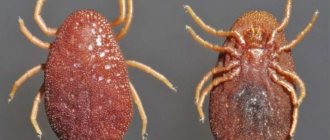
Sarcoptoid mites
They have sizes from 0.2 to 0.5 mm. There are different types of these parasites. They feed on blood, lymph, and dead epithelial cells. More often they affect large and small animals. They are known to veterinarians as the causative agents of scabies. When they come into contact with humans, they cause sarcoptoidosis, which is milder in humans than in animals. This is explained by the fact that man is a nonspecific host.
Sarcoptoidosis is milder in humans than in animals
Just like scabies, sarcoptoids dig tunnels in the epidermis of animals. When the mite gets to a person from an infected mammal, it causes pseudoscabies. It is accompanied by itching and redness of the epidermis, but the tick does not bite into the skin: conditions for reproduction are not suitable for it. Therefore, arthropods leave humans, and the symptoms of the disease go away on their own without treatment.
Sarcoptoid mites can appear in humans after contact with an infected animal, most often a dog.
There is a high risk of infection among livestock farmers caring for cattle, pigs and sheep. The palms, arms, and chest are most often affected. The skin turns red, a papular rash and itching appears. These symptoms go away on their own after some time. Those who have recovered from the disease develop hypersensitivity to ticks, which manifests itself as a periodic rash.
Recommendations and useful tips for treatment
To achieve more visible treatment results, dermatologists advise following the following recommendations:
- Cleanse the skin daily with special preparations that eliminate excessive oily sheen;
- Do not use hot water for washing , this procedure disrupts the natural protective functions of the skin;
- Reduce skin touching , especially in the facial area. A person’s hands may contain a large number of bacteria that contribute to the disruption of the structure of the skin and can cause the development of mites;
- Ensure the cleanliness of personal hygiene products and items for applying cosmetics;
- Avoid prolonged exposure to direct sunlight;
- Replace the feather pillow with padding polyester , very often down can cause a reaction and provoke the occurrence of demodex;
- Do not apply cosmetics in large quantities ; this method of applying cosmetics leads to clogged pores and the development of skin mites.
Experts recommend following such facial care methods even in the absence of tick infestation, as well as during treatment with medications.
The benefits and harm caused by Demodex
By feeding on the epithelial cells of the hair follicles and sebaceous glands, eating the secretions of the glands, demodex mites violate the integrity of the skin, this leads to the appearance of inflammatory infiltrates and pigmentation. The amount of tick metabolic products increases, toxins are released, and an entry point for infection is formed.
Thus, Demodex mites play the role of a mechanical and chemical irritant in the development of pathological changes in the skin, serving as carriers of microbes and viruses deep into the sebaceous glands of the hair follicles. Skin health suffers, pores expand, skin oiliness increases, skin becomes rough, red, and pimples appear.
Some medical sources believe that the demodex skin mite helps in clearing the epidermis of dead skin cells by eating it, which is clearly visible in the photo above.
Benefit
Despite the obvious harm, many talk about the benefits brought by ticks. Thus, foreign experts have proven through experiments that the Demodex mite is involved in the regulation of skin ph.
It also destroys a large number of microorganisms that fall on the surface of the skin. Demodex is the orderly of our skin, as it feeds on pieces of the epidermis with dead cells that have already sloughed off. Mites ferment fat and eat its excess - read about this in detail here - demodicosis treatment on the face - learn a lot of unexpected things about demodex.
Special diet
When dealing with demodex disease, experts often advise following a certain type of diet.
First of all, you should exclude:
- Salty food;
- Fatty foods;
- Fried and spicy foods;
- Smoked meats and sausages;
- Sweets;
- Canned and fatty meats.
During the treatment period, it is recommended to focus on the following fruits:
- Apples;
- Pears;
- Banana;
- Grapefruit (small quantity)
- Pomegranate;
- Kiwi.
Consumption of citrus fruits is not recommended, as this can contribute to the development of allergic reactions and aggravate the disease.
It is also recommended to introduce fermented milk products and whole grain cereals into the diet. Saturating the body with foods that contain fiber can reduce the deposition of toxins and wastes and improve natural metabolic processes.
The best drugs
As already mentioned, the modern drug market has a wide range of different drugs both for the destruction of subcutaneous mites and for eliminating the consequences of their activity. The most commonly used drugs are:
- Ointment "YAM" , which is a veterinary drug, but is prescribed in some cases for humans. The product is applied liberally to the face before bed for 10 days, every morning it must be thoroughly washed off using soap. After a month, a similar course will need to be repeated.
- Twice a day you can make lotions from a solution based on buckthorn bark ; they must be combined with the application of birch tar, which is then washed off with water and soap. Such measures are not a complete treatment and cannot replace pharmacological drugs, but will be a good addition to the main course.
- Suprastin and tavegil will help reduce the allergic reaction and relieve irritation caused by the activity of subcutaneous mites.
- Trichopolum is a tablet with antibacterial properties; a weekly course of administration is usually prescribed.
- Demodex Complex includes 10 different drugs designed to destroy subcutaneous mites and eliminate the symptoms caused by them. The set includes ointments, tonic for washing, medicinal tea and antiparasitic hair products.
Undergoing treatment and taking pharmacological drugs can cause the following consequences, which should not be feared:
- Peeling of the skin , which will indicate the death of parasites. The husk will fall off on its own; it should not be peeled off or treated with any means, as this may cause recovery processes in the mites.
- The appearance of new pimples is also a sign that the treatment is successful. They arise due to the death of deposited larvae and it is strictly forbidden to touch or squeeze out such pimples, since infection can cause a new surge in parasite activity.
Reviews
Reviews about the treatment of skin mites:
The role of iron mites in the development of skin diseases
The parasitism of these mites on humans can often be asymptomatic. On average, up to 55% of people are carriers of iron mites. With age, tick infestation increases and, according to some authors, in older people it reaches 100%. Because of this, their role in the development of skin diseases is assessed ambiguously, but it has been proven that the number of D. folliculorum
more than 5 copies per sq. cm.
The number of iron mites significantly increases in patients with some forms of rosacea: mites of the genus Demodex
to be involved in the pathogenesis of these diseases.
1.4
Prevention measures
To prevent the occurrence of such an unpleasant disease as skin mites, it is recommended to follow the following prevention methods:
- Change bed linen regularly;
- Have a separate personal towel for your face;
- Do not use other people's personal hygiene products;
- Maintain proper nutrition, exercise outdoors;
- Treat diseases of internal organs in a timely manner;
- Take additional vitamin complexes;
- After treatment with antibiotics, use special drugs to restore the immune system;
- Use sunscreen when exposed to direct sunlight for long periods of time.
Regular adherence to prevention methods can eliminate the likelihood of tick infection.
How is demodicosis transmitted?
Considering the routes of transmission and wondering how Demodex is transmitted from person to person, you should pay attention to the fact that the household route is quite common in this disease. Therefore, you should not share things and hygiene items with the patient. Not least important is the contact type, for example, the contact of eyelashes, eyebrows, affected skin with healthy ones. Therefore, Demodex can also be infected through sexual contact. A sick mother can transmit the infection to her child during childbirth or during feeding, but the disease manifests itself only a week after birth.
At the same time, there is no clear answer whether demodex is contagious or not, since this microorganism is an opportunistic human flora and lives in the body of 90-95% of the world's inhabitants. Even if the parasite has entered the body of a healthy person, it may not cause illness if there are no accompanying factors, such as decreased immunity, hormonal and metabolic disruptions.
In addition, Demodex can live on the body of pets. It will not cause any inconvenience to the animal, but it can pass on to its owner. There is also a genetic connection between patients. Less commonly, infection occurs due to consumption of junk food and alcohol during inflammatory processes in the digestive tract.
Clinical manifestations
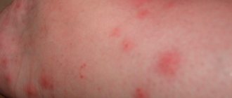
Have you suddenly noticed increased skin roughness? Demodectic mange should not be ruled out. There may also be other symptoms:
- skin itching or peeling;
- redness;
- increased skin sensitivity;
- burning sensation of the face;
- The skin felt rough, like sandpaper.
Depending on the location of the lesion, the following types of disease are distinguished:
- Eye shape. Demodicosis of the eyelids is accompanied by itching, pain, and blepharitis. The eyes quickly get tired, the eyelids swell, and the eyelashes fall out. There may be inflammation of the mucous membrane of the eyes - conjunctivitis.
- Damage to hairy areas. Irritation, itching, and peeling of the skin appear. The hairline becomes sparse and then the hair shafts fall out completely.
- Damage to the porioral and ear areas. The skin becomes rough and itchy.
Then there may be signs of dermatitis: redness, irritation, swelling of the tissue. Based on skin manifestations, two types of demodicosis are distinguished:
- Erythematous. It is characterized by the appearance of red spots on the face a week before the full picture of the disease. It resembles a mosquito bite that is a little itchy and itchy. Then the spots increase in size and the skin becomes oily.
- Papular-pustular. Against the background of redness and irritation of the skin, pustules appear. Gradually they grow, the surface of the dermis becomes uneven, bumpy, painful and itchy. The peculiarity of demodicosis is that the pustules are located nearby in the form of a chain. Itching is most pronounced at night during the parasite's breeding season.
Demodicosis can be combined with other pathologies, aggravating their course. Let's look at the main ones that occur most often.
Rosacea
Numerous studies have reported increased levels of parasites in patients with rosacea. Human demodicosis can manifest as a dry type of rosacea and is called rosacea-like demodicosis.
Let's look at the main differences
| Rosacea caused by Demodex | Common rosacea |
| Characterized by dryness, flaking of follicles, superficial vesicles and pustules | Characterized by oily skin, lack of scales and deeper skin lesions |
| Full recovery after treatment | Doesn't go all the way |
Demodectic rosacea completely goes away after the therapy prescribed by the doctor.
If you do not wash your face or oversaturate the skin with oil creams, then demodicosis on the face intensifies. This is due to the fact that ticks feed on lipids and begin to actively reproduce. In addition, the sebaceous ducts become clogged and rashes appear.
Dermatitis
With dermatitis, itching appears on the face, there may be redness, rash, papules, pustules, acne, comedones. In some people, dermatitis may present as a dry, flaky, rough face without papules or pustules.
Steroid dermatitis and rosacea
Appears when using corticosteroid drugs. Due to their use, local immunity decreases, and D. Folliculorum begins to actively reproduce.
Androgenetic alopecia
Mites can contribute to baldness. In the affected area, inflammatory processes around the sebaceous glands are activated, followed by the development of fibrous tissue and changes in hormonal levels. The sebaceous glands increase in size, producing large amounts of lipids. Hair follicles become depleted and hair stops growing.
Madaroz
This is what is called eyelash loss. The mite penetrates the follicles on the eyelids, causing inflammation and swelling in them. Eyelashes become thin and brittle and then fall out.
Trigger mechanism of demodicosis disease
Scabies and glandular acne are common skin parasites, the mechanism of activation of which is not fully understood. To this day, it is unclear why microscopic mites of this nature begin to multiply rapidly at a certain moment and suddenly stop multiplying under similar conditions.
Experts consider the use of cosmetics containing hormonal components to be one of the main triggers for demodicosis. The latter are the favorite food of parasitic mites.
In such situations, the pathogens of the disease not only begin to show increased activity on the skin, but also end up in containers with cosmetics, where they also develop and then re-infect a person.
Another trigger is deterioration of the skin condition and the appearance of looseness in its structure. In most cases, the problem affects women. Under such conditions, damage to the epidermis by the causative agent of the disease in men occurs much less frequently. The reason is frequent shaving, during which the blade removes dead, keratinized layers of skin where the microscopic mite has barely managed to gain a foothold.
It is possible to defeat parasites!
Antiparasitic Complex® - Reliable and safe removal of parasites in 21 days!
- The composition includes only natural ingredients;
- Does not cause side effects;
- Absolutely safe;
- Protects the liver, heart, lungs, stomach, skin from parasites;
- Removes waste products of parasites from the body.
- Effectively destroys most types of helminths in 21 days.
There is now a preferential program for free packaging. Read expert opinion.
Read further:
Main types of streptococci: description and life cycle of development
Demodex brevis: structure of the parasite, diagnosis and how to treat
Life cycle of development of the human roundworm, stages of development of the parasite in the body
Tumbu fly (photo): description of the parasite, life cycle of larval development
Dermatobia Hominis: description, development cycle, symptoms and treatment
Taenia saginata: description of the parasite, life cycle of development, routes of infection and treatment
First aid for a tick bite
First aid for a tick bite involves timely detection and proper removal of the parasite . If you remove a tick correctly using the instructions described above, the likelihood of contracting encephalitis, borreliosis and other viral diseases is reduced to almost zero, even if the insect was infected.
Virologists do not recommend emergency prevention, especially in field conditions far from medical centers. The problem is the pronounced side effects of emergency prophylactic drugs. Taking immunoglobulin (tick-borne encephalitis) or doxycycline (borreliosis), a person feels general fatigue, nausea, severe muscle pain and headaches, even fainting. Convulsions may occur.
Therefore, the best thing a person who has been bitten by a tick can do is to remove it correctly and immediately go to the nearest medical center. point, taking with you the extracted parasite . Doctors will take the insect for examination and, based on the analysis results, will be able to determine what viruses the tick was infected with. This approach gives the most effective results because:
- Doctors learn the patient’s possible diagnosis at an early stage, which greatly influences the effectiveness of future treatment. It is much easier to fight viruses in the early stages .
- A person who goes to a medical facility is already under the supervision of doctors.
- Ticks often carry multiple viruses at the same time. Laboratory studies of the insect will allow us to identify all the viruses that the tick carries and develop relevant drug treatment.
Important! Every tick bite is potentially dangerous. Therefore, it is better to go to the hospital. This is especially true for residents of areas endemic for tick-borne encephalitis and borreliosis.
Mechanisms of action of iron mites on humans
The impact of iron mites on the human body is multifaceted.
1. When dust mites feed, mechanical destruction of the cell walls of the follicles and sebaceous glands occurs, which contributes to cell destruction, keratinization, pigmentation and the formation of granulomas and inflammatory infiltrates.
2. There is an assumption that iron mites contribute to the transmission of bacteria. Staphylococci were found on the surface of the ticks' bodies and in their digestive system, and they were also detected on the eyelashes affected by ticks in 69% of cases. True, staphylococcus can also be inoculated from eyelashes that are not affected by mites, and sometimes inoculation of the contents of pustules with rosacea turns out to be sterile.
3. Iron mites, parasitizing a person, apparently suppress his immune system, which allows the mites themselves and opportunistic microorganisms to successfully colonize the host’s skin.
4. Recently, the number of patients with a genetic predisposition to atopy has been steadily increasing. The number of patients with hypersensitivity to house dust mites (family Pyroglyphidae
). It is possible that iron mites and house dust mites have common antigenic structures. It is possible that the sensitizing properties of iron mites (their bodies and metabolites) are underestimated.
5. Some patients, especially those who have problems with their facial skin, develop acarophobia after they learn about the discovery of iron mites, which causes various psychological difficulties.
1.6